WBR0188
| Author | [[PageAuthor::Ogheneochuko Ajari, MB.BS, MS [1] (Reviewed by Will Gibson)]] |
|---|---|
| Exam Type | ExamType::USMLE Step 1 |
| Main Category | MainCategory::Microbiology |
| Sub Category | SubCategory::Infectious Disease |
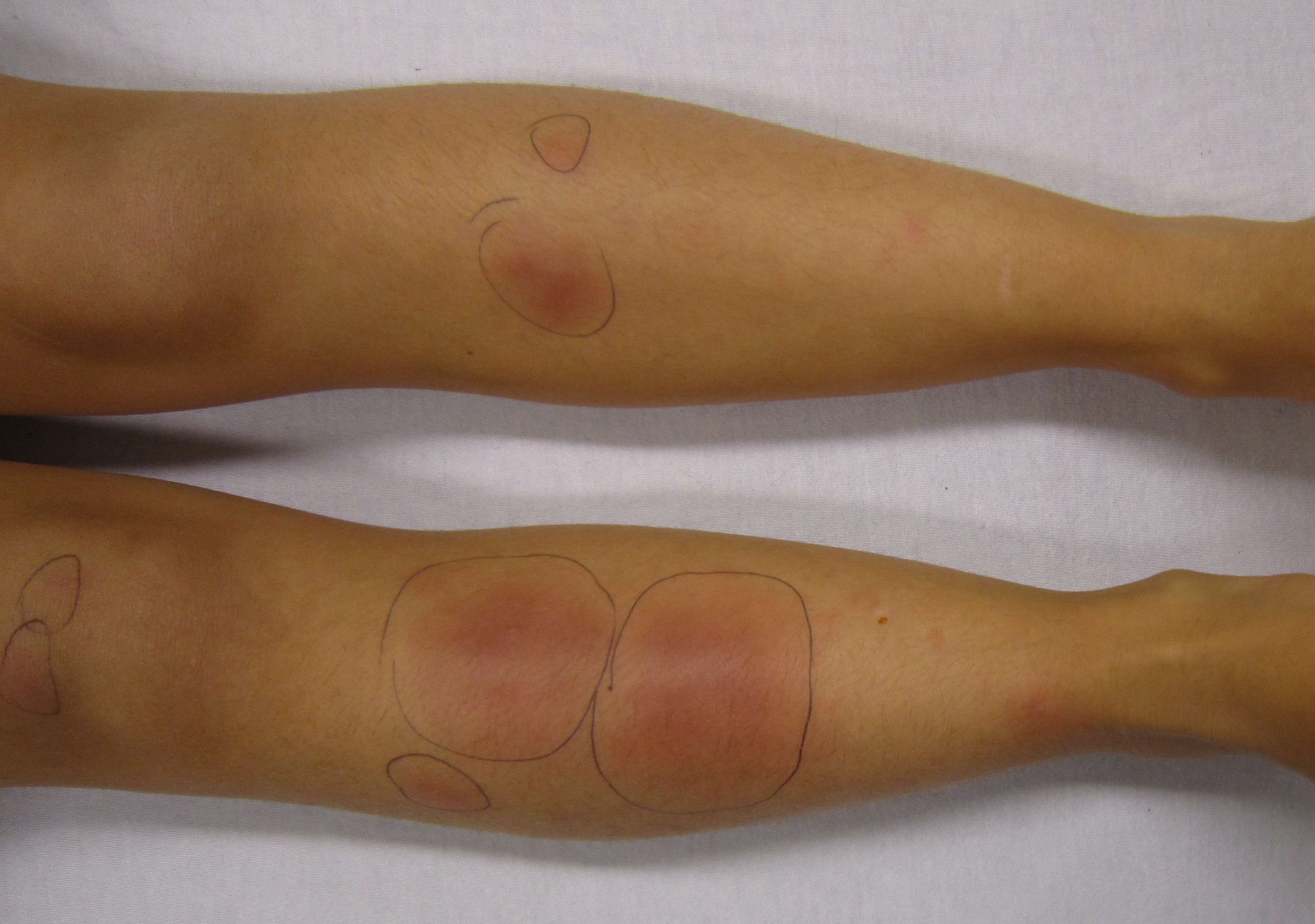
| Prompt | [[Prompt::A 65-year old woman with a history of rheumatoid arthritis presents to urgent care for fatigue, myalgias, cough and fever. She reports a forty year smoking history, but has no otherwise remarkable past medical history. She emigrated from Mexico as a teenager and has been a migrant worker in southern California since. Three weeks ago, her home was destroyed in an earthquake and she has been living with her son. Physical exam reveals an erythematous rash on the lower limbs (pictured below). A chest radiograph reveals multiple nodules and hilar adenopathy. Which of the following is most likely to be seen on microscopic examination of a lung tissue biopsy? |
| Answer A | AnswerA::Broad based budding yeast |
| Answer A Explanation | AnswerAExp::Broad based budding yeast is seen in Blastomyces dermatitidis. |
| Answer B | AnswerB::Spherules with endospores |
| Answer B Explanation | AnswerBExp::Spherules with endospores characteristically describes Coccidioides immitis. |
| Answer C | AnswerC::Septate hyphae branching dichotomously at acute angles |
| Answer C Explanation | AnswerCExp::Septate hyphae branching dichotomously at acute angles describes Aspergillus fumigatus. |
| Answer D | AnswerD::Non septate hyphae with broad angles |
| Answer D Explanation | AnswerDExp::Non septate hyphae with broad angles describes Mucor species. |
| Answer E | AnswerE::Monomorphic encapsulated yeast |
| Answer E Explanation | AnswerEExp::Monomorphic encapsulated yeast describes Cryptococcus neoformans. |
| Right Answer | RightAnswer::B |
| Explanation | [[Explanation::The patient in this vignette has developed symptoms of systemic coccidiomycosis. Coccidiodes immitis is a pathogenic fungus that is endemic to the southwestern United States. It is most often acquired through inhalation of a spore. In extremely rare cases, it can be acquired through a spore entering an open wound in the skin. Most people (60%) do not develop any symptoms of infection, the remaining 40% tend to experience a mild pneumonia. In 5-10% of cases, patients may develop more aggressive pulmonary disease, or chronic infection. These patients are often immunocompromised and sometimes develop systemic disease (1% of all patients).
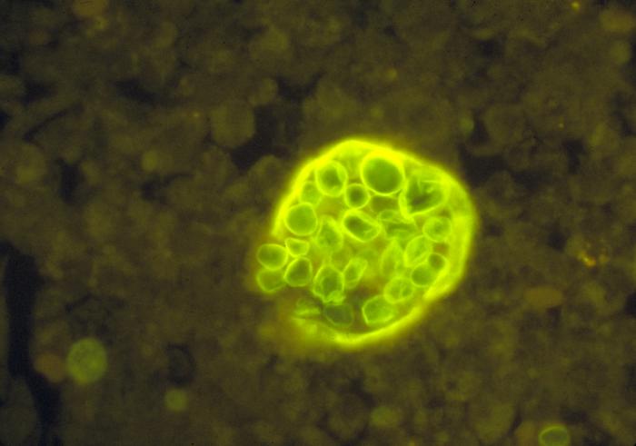
Pulmonary coccidiomycosis usually presents with fatigue, cough, myalgias, fever and night sweats. Most patients' symptoms will resolve without medical intervention. Systemic coccidiomycosis can manifest with some of the additional symptoms seen in this patient such as rash (usually erythema nodosum or erythema multiforme). The different systemic mycoses can be difficult to distinguish based on symptoms alone, as they all primarily cause pulmonary disease. However, several factors make Coccidiodes the most likely etiologic agent in this case: (i). The patient comes from the southwestern United States, an endemic area of coccidiodes (see figure below). (ii). She comes to medical attention shortly after an earthquake. The incidence of coccidiomycosis tends to increase after earthquakes as spores are released from the earth and inhaled. (iii) The patient has rheumatoid arthritis and likely takes medications such as methotrexate that cause immunocompromization. She is therefore, more susceptible to coccidiodes infection. (iv) Skin rash is more rare in other systemic mycoses. Given that the patient has been infected with Coccidiodes immitis, the question now asks for the microscopic appearance of the organism. The diagnostic form of C. immitis in tissue is a spherule with endospores (pictured below).

First Aid 2014 page 146]] |
| Approved | Approved::Yes |
| Keyword | WBRKeyword::Microbiology, WBRKeyword::Eukaryotes, WBRKeyword::Yeast, WBRKeyword::Coccidioidomycosis, WBRKeyword::C immitis, WBRKeyword::Coccidioides immitis, WBRKeyword::Valley fever |
| Linked Question | Linked:: |
| Order in Linked Questions | LinkedOrder:: |

