Babesiosis laboratory findings
Editor-In-Chief: C. Michael Gibson, M.S., M.D. [1]
|
Babesiosis Microchapters |
|
Diagnosis |
|---|
|
Treatment |
|
Case Studies |
|
Babesiosis laboratory findings On the Web |
|
American Roentgen Ray Society Images of Babesiosis laboratory findings |
|
Risk calculators and risk factors for Babesiosis laboratory findings |
Overview
Babesiosis is easy to diagnose but only if it is suspected. It will not show up on any routine tests. It must be suspected when a persons with exposure in an endemic area develops persistent fevers and hemolytic anemia.
Laboratory Findings
Electrolyte and Biomarker Studies
Babesiosis can be diagnosed by direct examination of the blood, with serology, or with PCR-based tests. Serologic testing for antibodies provides a supporting evidence for a potential infection. Other laboratory findings include decreased numbers of red blood cells and platelets on complete blood count.
Microscopy
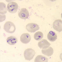
In symptomatic people, babesiosis usually is diagnosed by examining blood specimens under a microscope and seeing Babesia parasites inside red blood cells. [1]

Gallery
-
Babesia microti in blood smear. Giemsa stain. Parasite. From Public Health Image Library (PHIL). [2]
-
Babesia microti in blood smear. Giemsa stain. Parasite. From Public Health Image Library (PHIL). [2]
-
Babesia microti in blood smear. Giemsa stain. Parasite. From Public Health Image Library (PHIL). [2]
-
Babesia microti in blood smear. Giemsa stain. Parasite. From Public Health Image Library (PHIL). [2]
-
Babesia microti in blood smear. Giemsa stain. Parasite. From Public Health Image Library (PHIL). [2]
-
Babesia microti in blood smear. Giemsa stain. Parasite. From Public Health Image Library (PHIL). [2]
-
Babesia microti in blood smear. Giemsa stain. Parasite. From Public Health Image Library (PHIL). [2]
-
Babesia microti in blood smear. Giemsa stain. Parasite. From Public Health Image Library (PHIL). [2]
-
This blood smear micrograph reveals a Babesia sp. tetrad formation. From Public Health Image Library (PHIL). [2]
-
This blood smear micrograph revealed the presence of Babesia sp. ring formations inside the host erythrocytes. From Public Health Image Library (PHIL). [2]
-
This photomicrograph revealed the presence of an "older ring-form" of a Babesia sp. protozoan parasite in a blood smear. This older ring-form was located within an erythrocyte, and was displaying two chromatin masses. From Public Health Image Library (PHIL). [2]
-
Blood smear showing large Babesia rings in erythrocytes. From Public Health Image Library (PHIL). [2]
-
At a magnification of 1000X, this blood smear photomicrograph revealed the presence of a number of intra-erythrocytic forms of Babesia sp. hemoprotozoan parasites. From Public Health Image Library (PHIL). [2]
-
Blood smear showing larger trophic stage of Babesia microti in erythrocyte. From Public Health Image Library (PHIL). [2]
-
Note the developmental “tetrad” configuration of these Babesia sp. trophozoites, which resemble P. falciparum. From Public Health Image Library (PHIL). [2]
![Babesia microti in blood smear. Giemsa stain. Parasite. From Public Health Image Library (PHIL). [2]](/images/0/08/Babesiosis15.jpeg)
![Babesia microti in blood smear. Giemsa stain. Parasite. From Public Health Image Library (PHIL). [2]](/images/5/54/Babesiosis14.jpeg)
![Babesia microti in blood smear. Giemsa stain. Parasite. From Public Health Image Library (PHIL). [2]](/images/1/12/Babesiosis13.jpeg)
![Babesia microti in blood smear. Giemsa stain. Parasite. From Public Health Image Library (PHIL). [2]](/images/3/3f/Babesiosis12.jpeg)
![Babesia microti in blood smear. Giemsa stain. Parasite. From Public Health Image Library (PHIL). [2]](/images/1/17/Babesiosis11.jpeg)
![Babesia microti in blood smear. Giemsa stain. Parasite. From Public Health Image Library (PHIL). [2]](/images/1/17/Babesiosis10.jpeg)
![Babesia microti in blood smear. Giemsa stain. Parasite. From Public Health Image Library (PHIL). [2]](/images/a/aa/Babesiosis09.jpeg)
![Babesia microti in blood smear. Giemsa stain. Parasite. From Public Health Image Library (PHIL). [2]](/images/d/d5/Babesiosis08.jpeg)
![This blood smear micrograph reveals a Babesia sp. tetrad formation. From Public Health Image Library (PHIL). [2]](/images/a/a6/Babesiosis07.jpeg)
![This blood smear micrograph revealed the presence of Babesia sp. ring formations inside the host erythrocytes. From Public Health Image Library (PHIL). [2]](/images/2/2b/Babesiosis06.jpeg)
![This photomicrograph revealed the presence of an "older ring-form" of a Babesia sp. protozoan parasite in a blood smear. This older ring-form was located within an erythrocyte, and was displaying two chromatin masses. From Public Health Image Library (PHIL). [2]](/images/0/0e/Babesiosis05.jpeg)
![Blood smear showing large Babesia rings in erythrocytes. From Public Health Image Library (PHIL). [2]](/images/6/64/Babesiosis04.jpeg)
![At a magnification of 1000X, this blood smear photomicrograph revealed the presence of a number of intra-erythrocytic forms of Babesia sp. hemoprotozoan parasites. From Public Health Image Library (PHIL). [2]](/images/9/98/Babesiosis03.jpeg)
![Blood smear showing larger trophic stage of Babesia microti in erythrocyte. From Public Health Image Library (PHIL). [2]](/images/d/d8/Babesiosis02.jpeg)
![Note the developmental “tetrad” configuration of these Babesia sp. trophozoites, which resemble P. falciparum. From Public Health Image Library (PHIL). [2]](/images/f/ff/Babesiosis01.jpeg)