WBR0438: Difference between revisions
Jump to navigation
Jump to search
Rim Halaby (talk | contribs) No edit summary |
No edit summary |
||
| Line 1: | Line 1: | ||
{{WBRQuestion | {{WBRQuestion | ||
|QuestionAuthor={{Rim}} | |QuestionAuthor={{Rim}}, {{AJL}} {{Alison}} | ||
|ExamType=USMLE Step 1 | |ExamType=USMLE Step 1 | ||
|MainCategory=Physiology | |MainCategory=Physiology | ||
| Line 20: | Line 20: | ||
|MainCategory=Physiology | |MainCategory=Physiology | ||
|SubCategory=Cardiology | |SubCategory=Cardiology | ||
|Prompt=A 73 year old | |Prompt=A 73-year-old male with hypertension presents to the emergency room with complaints of shortness of breath at rest. Upon evaluation, you observe that his ventricles are not appropriately relaxing and filling, leading you to diagnose the patient with heart failure. Which of the following phases on electrocardiography (ECG) scheme (illustrated below) represents ventricular relaxing and filling? | ||
[[Image:Normal ECG.png|400px]] | [[Image:Normal ECG.png|400px]] | ||
|Explanation= | |Explanation=Upon [[ECG]], waves represent [[depolarization]] and [[repolarization]] effects, while baseline represents the absence of net depolarization or repolarization, frequently observed in cases of muscular rest or contraction. On ECG, [[P wave]]s represent [[atrial depolarization]], [[PR segment]]s represent [[AV nodal delay]], [[QRS complex]]es represent [[ventricular depolarization]] and simultaneous [[atrial repolarization]], [[ST segment]] represents time during which [[ventricular]] contraction is occurring, [[T wave]] represents [[ventricular repolarization]], and [[TP interval]]s represent the time during which ventricles relax and refill. | ||
|EducationalObjectives= On ECG, [[TP interval]]s represent ventricles relaxing and refilling. | |||
|AnswerA=PR | |References= Berne RM. Cardiovascular physiology. Annu. Rev. Physiol. 1981;43:357-358 | ||
|AnswerAExp=PR | |||
|AnswerB=ST | |AnswerA=PR segments | ||
|AnswerBExp=ST | |AnswerAExp=PR segments represent AV nodal delay. | ||
|AnswerC=QRS | |AnswerB=ST segments | ||
|AnswerCExp=QRS | |AnswerBExp=ST segments represent ventricle contraction. | ||
|AnswerD=QT | |AnswerC=QRS complexes | ||
|AnswerDExp=QT | |AnswerCExp=QRS complexes represent ventricular depolarization and simultaneous atrial repolarization. | ||
|AnswerE=TP | |AnswerD=QT intervals | ||
|AnswerEExp=TP | |AnswerDExp=QT intervals represent total duration of ventricular depolarization and repolarization. | ||
|AnswerE=TP intervals | |||
|AnswerEExp=TP intervals represent ventricular relaxation and refilling. | |||
|RightAnswer=E | |RightAnswer=E | ||
|WBRKeyword=ECG, electrocardiogram, ventricle, filling, relaxation, wave, repolarization | |WBRKeyword=ECG, electrocardiogram, ventricle, filling, relaxation, wave, repolarization, cardiology, cardiovascular, | ||
|Approved= | |Approved=Yes | ||
}} | }} | ||
Revision as of 19:02, 22 July 2014
| Author | [[PageAuthor::Rim Halaby, M.D. [1], Alison Leibowitz [2] (Reviewed by Alison Leibowitz)]] |
|---|---|
| Exam Type | ExamType::USMLE Step 1 |
| Main Category | MainCategory::Physiology |
| Sub Category | SubCategory::Cardiology |
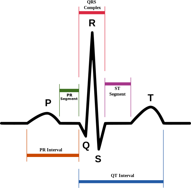
| Prompt | [[Prompt::A 73-year-old male with hypertension presents to the emergency room with complaints of shortness of breath at rest. Upon evaluation, you observe that his ventricles are not appropriately relaxing and filling, leading you to diagnose the patient with heart failure. Which of the following phases on electrocardiography (ECG) scheme (illustrated below) represents ventricular relaxing and filling? |
| Answer A | AnswerA::PR segments |
| Answer A Explanation | AnswerAExp::PR segments represent AV nodal delay. |
| Answer B | AnswerB::ST segments |
| Answer B Explanation | AnswerBExp::ST segments represent ventricle contraction. |
| Answer C | AnswerC::QRS complexes |
| Answer C Explanation | AnswerCExp::QRS complexes represent ventricular depolarization and simultaneous atrial repolarization. |
| Answer D | AnswerD::QT intervals |
| Answer D Explanation | AnswerDExp::QT intervals represent total duration of ventricular depolarization and repolarization. |
| Answer E | AnswerE::TP intervals |
| Answer E Explanation | AnswerEExp::TP intervals represent ventricular relaxation and refilling. |
| Right Answer | RightAnswer::E |
| Explanation | [[Explanation::Upon ECG, waves represent depolarization and repolarization effects, while baseline represents the absence of net depolarization or repolarization, frequently observed in cases of muscular rest or contraction. On ECG, P waves represent atrial depolarization, PR segments represent AV nodal delay, QRS complexes represent ventricular depolarization and simultaneous atrial repolarization, ST segment represents time during which ventricular contraction is occurring, T wave represents ventricular repolarization, and TP intervals represent the time during which ventricles relax and refill. Educational Objective: On ECG, TP intervals represent ventricles relaxing and refilling. |
| Approved | Approved::Yes |
| Keyword | WBRKeyword::ECG, WBRKeyword::electrocardiogram, WBRKeyword::ventricle, WBRKeyword::filling, WBRKeyword::relaxation, WBRKeyword::wave, WBRKeyword::repolarization, WBRKeyword::cardiology, WBRKeyword::cardiovascular |
| Linked Question | Linked:: |
| Order in Linked Questions | LinkedOrder:: |
