Atrioventricular block electrocardiogram: Difference between revisions
(Created page with "__NOTOC__ {{Atrioventricular block}} {{CMG}} == Overview == == Electrocardiogram == <div align="left"> <gallery heights="225" widths="225"> Image:LAE.jpg|First degree AV blo...") |
|||
| Line 4: | Line 4: | ||
== Overview == | == Overview == | ||
== Electrocardiogram == | == Electrocardiogram Findings == | ||
<div align="left"> | <div align="left"> | ||
| Line 12: | Line 12: | ||
</gallery> | </gallery> | ||
</div> | </div> | ||
---- | |||
Shown below is an EKG showing sinus rhythm with a prolonged pr interval (>120ms.) which is first degree A/V block. There is also a left axis deviation (axis between -30 and -90 degrees) with r waves in the inferior leads. This axis deviation is consitent with a left anterior fasicular block. | |||
[[File:AVBlockEKG.jpg|center|500px]] | |||
---- | |||
==Sources== | |||
Copyleft images obtained courtesy of ECGpedia, http://en.ecgpedia.org/index.php?title=Special:NewFiles&dir=prev&offset=20080806182927&limit=500 | |||
==References== | ==References== | ||
Revision as of 15:51, 15 October 2012
|
Atrioventricular block Microchapters |
|
Diagnosis |
|---|
|
Treatment |
|
Case Studies |
|
Atrioventricular block electrocardiogram On the Web |
|
American Roentgen Ray Society Images of Atrioventricular block electrocardiogram |
|
Risk calculators and risk factors for Atrioventricular block electrocardiogram |
Editor-In-Chief: C. Michael Gibson, M.S., M.D. [1]
Overview
Electrocardiogram Findings
-

First degree AV block is a misnomer in that every P wave is conducted to the ventricles, however, with a PR interval exceeding 200 msec. Prolonged PR conduction, a more appropriate classification for this conduction disturbance, may be the result of conduction delay within the atrium, AV node, bundle of His or bundle branches. Prolongation of the PR interval most often indicates AV nodal conduction delay.
-
Two-to-one AV block can represent benign block within the AV node or disease of the His-Purkinje system. Certain electrocardiographic features and maneuvers can help in distinguishing where the location of block exists. A long PR interval with a narrow QRS suggests an intranodal block. A short PR interval with intraventricular conduction delay or bundle branch block suggests disease below the node. Responses to atropine, exercise and carotid sinus massage can be helpful in diagnosis. Atropine will improve AV nodal conduction but will worsen block within diseased His-Purkinje fibers. Exercise has a similar effect, improving conduction in cases where block exists only in the node, but worsening when block is subnodal. Alternatively, Carotid Sinus Massage will slow conduction when block occurs in the AV node, but will improve conduction in diseased His-Purkinje tissue by allowing for refractoriness to recover


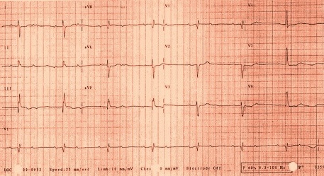
Shown below is an EKG showing sinus rhythm with a prolonged pr interval (>120ms.) which is first degree A/V block. There is also a left axis deviation (axis between -30 and -90 degrees) with r waves in the inferior leads. This axis deviation is consitent with a left anterior fasicular block.

Sources
Copyleft images obtained courtesy of ECGpedia, http://en.ecgpedia.org/index.php?title=Special:NewFiles&dir=prev&offset=20080806182927&limit=500