Syphilis physical examination: Difference between revisions
Jump to navigation
Jump to search
No edit summary |
|||
| Line 1: | Line 1: | ||
Syphilis is a curable [[sexually transmitted disease]] caused by the ''[[Treponema pallidum]]'' [[spirochete]]. The route of transmission of syphilis is almost always by [[sexual]] contact, although there are examples of [[congenital syphilis]] via transmission from mother to child [[in utero]]. The signs and [[symptoms]] of syphilis are numerous; before the advent of [[serological testing]], precise [[diagnosis]] was very difficult. | Syphilis is a curable [[sexually transmitted disease]] caused by the ''[[Treponema pallidum]]'' [[spirochete]]. The route of transmission of syphilis is almost always by [[sexual]] contact, although there are examples of [[congenital syphilis]] via transmission from mother to child [[in utero]]. The signs and [[symptoms]] of syphilis are numerous; before the advent of [[serological testing]], precise [[diagnosis]] was very difficult. | ||
Revision as of 16:24, 14 March 2012
Syphilis is a curable sexually transmitted disease caused by the Treponema pallidum spirochete. The route of transmission of syphilis is almost always by sexual contact, although there are examples of congenital syphilis via transmission from mother to child in utero. The signs and symptoms of syphilis are numerous; before the advent of serological testing, precise diagnosis was very difficult.
Physical examination
Primary syphilis: Chancre
- Afebrile
- Chancre:
- single painless papule which rapidly progresses an ulcerated, indurated lesion with a surrounding red areola
- usually located on the penis,cervix, labia, anal canal, rectum or oral cavity
- highly infectious lesion
- Regional lymphadenopathy accompanies primary lesion.
- onset within a week
- unilateral or bilateral
- lymph nodes are firm, painless, non-tender and non-suppurative
- Primary chancre heals spontaneously within 4-6weeks; however, regional lymphadenopathy may persist for longer periods.
Secondary syphilis: Condylomata Lata
- Develops 6-8 weeks after the appearance of primary chancre.
- Cardinal signs include:
- Skin rash: initial macular lesions on the trunk and proximal limbs with progressive generalized papular rash and may cause necrotic ulcers.
- Lymphadenopathy: localized or generalized, firm and non-tender
- Condylomata lata:
- reddish-brown papular lesions on the intertriginous areas that coalesce and enlarge into large plaques known as condylomata lata
- lesions usually progress from painful vesicular pattern to erosive lesions with resultant broad, grey-white highly infectious lesions
- Superficial mucosal patches:
- painless
- may be macular, papular, pustular or mixed
- located on the palate, pharynx, larynx, penis, vulva, anal canal or rectum
-
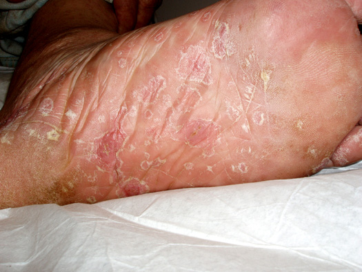
Erruption on Sole of Foot Associated with Secondary Syphilis.
-
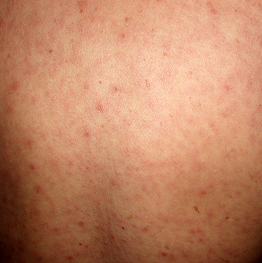
Generalized (Maculo-Papular) Eruption Associated with Secondary Syphilis.
-
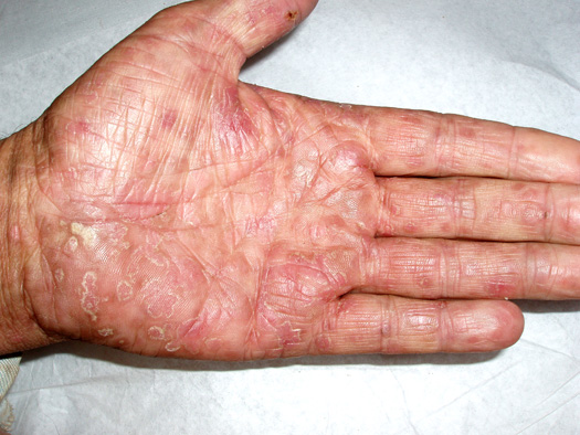
Palmar Erruption Associated with Secondary Syphilis.
-
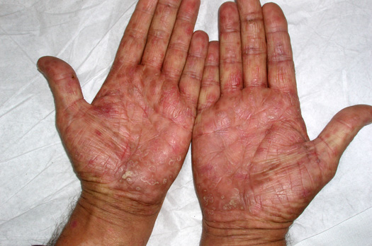
Palmar Erruption Associated with Secondary Syphilis.


Tertiary syphilis: Gumma
- soft, asymmetric, coalscent granulomatous lesion
- solitary lesions less than a centimeter in diameter
- appear almost anywhere in the body including in the skeleton
- cutaneous gumma: indurated, nodular, papulosquamous to ulcerative lesions with peripheral hyperpigmentation
- Cardiovascular manifestation secondary to aortic dilation with resultant aortic regurgitation:
- diastolic murmur
- de Musset's sign[1] a bobbing of the head that de Musset first noted in Parisian prostitutes
- Neurological manifestation:
- Asymptomatic meningitis
- Asymptomatic neurosyphilis usually has no signs or symptoms and is diagnosed exclusively with the presence of CSF abnormalities notably pleocytosis, elevated protein, decreased glucose or a positive VDRL test.
- Symptomatic meningitis
- develops within 6-months to several years of primary infection
- typical meningitis symptoms present
- cranial nerve abnormalities may be observed
- Meningovascular syphilis
- occurs a few months to 10 years (average, 7 years) after the primary infection
- associated with prodromal symptoms lasting weeks to months before focal deficits are identifiable
- focal deficits initially are intermittent or progress slowly over a few days
- clinical present with CNS vascular insufficiency or stroke involving the middle cerebral artery
- Parenchymatous neurosyphilis
- develops 15-20 years after primary infection
- clinical presents as general paresis or tabes dorsalis with resultant ataxia
- argyll robertson pupil: small irregular pupil
Ophthalmic examination
- Slit-lamp examination and ophthalmic examination may be helpful to differentiate between acquired and congenital syphilis.
- Presence of interstitial keratitis is suggestive of congenital syphilis with latent infection of unknown duration.
Clinical pearl: Syphilis detecting Handshake
{{#ev:youtube|SAedwyzTMWA}}