Renal papillary necrosis CT: Difference between revisions
Jump to navigation
Jump to search
No edit summary |
mNo edit summary |
||
| Line 11: | Line 11: | ||
** Findings on [[CT urography]] suggestive of [[renal papillary necrosis]] include multiple small accumulations of [[contrast media]] in the [[papillary]] regions. <ref name="pmid28076022">{{cite journal| author=Kawamoto S, Duggan P, Sheth S, Miyamoto H, Kazi ZN, Fishman EK| title=Renal Papillary and Calyceal Lesions at CT Urography: Genitourinary Imaging. | journal=Radiographics | year= 2017 | volume= 37 | issue= 1 | pages= 358-359 | pmid=28076022 | doi=10.1148/rg.2017160089 | pmc= | url=https://www.ncbi.nlm.nih.gov/entrez/eutils/elink.fcgi?dbfrom=pubmed&tool=sumsearch.org/cite&retmode=ref&cmd=prlinks&id=28076022 }} </ref> | ** Findings on [[CT urography]] suggestive of [[renal papillary necrosis]] include multiple small accumulations of [[contrast media]] in the [[papillary]] regions. <ref name="pmid28076022">{{cite journal| author=Kawamoto S, Duggan P, Sheth S, Miyamoto H, Kazi ZN, Fishman EK| title=Renal Papillary and Calyceal Lesions at CT Urography: Genitourinary Imaging. | journal=Radiographics | year= 2017 | volume= 37 | issue= 1 | pages= 358-359 | pmid=28076022 | doi=10.1148/rg.2017160089 | pmc= | url=https://www.ncbi.nlm.nih.gov/entrez/eutils/elink.fcgi?dbfrom=pubmed&tool=sumsearch.org/cite&retmode=ref&cmd=prlinks&id=28076022 }} </ref> | ||
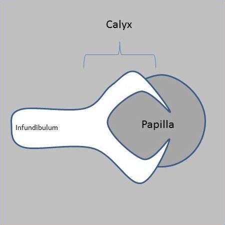
** The lobster claw sign is a diagnostic finding on [[CT]] [[urography]] in the papillary subtype of [[renal papillary necrosis]] that develops due to elongation of the [[minor calyx]] fornices following [[ischemia]] and destruction of the [[renal]] [[papilla]]. <ref name="pmid30309154">{{cite journal| author=Xiang H, Han J, Ridley WE, Ridley LJ| title=Lobster claw sign: Renal papillary necrosis. | journal=J Med Imaging Radiat Oncol | year= 2018 | volume= 62 Suppl 1 | issue= | pages= 90 | pmid=30309154 | doi=10.1111/1754-9485.37_12784 | pmc= | url=https://www.ncbi.nlm.nih.gov/entrez/eutils/elink.fcgi?dbfrom=pubmed&tool=sumsearch.org/cite&retmode=ref&cmd=prlinks&id=30309154 }} </ref> | ** The lobster claw sign is a diagnostic finding on [[CT]] [[urography]] in the papillary subtype of [[renal papillary necrosis]] that develops due to elongation of the [[minor calyx]] fornices following [[ischemia]] and destruction of the [[renal]] [[papilla]]. <ref name="pmid30309154">{{cite journal| author=Xiang H, Han J, Ridley WE, Ridley LJ| title=Lobster claw sign: Renal papillary necrosis. | journal=J Med Imaging Radiat Oncol | year= 2018 | volume= 62 Suppl 1 | issue= | pages= 90 | pmid=30309154 | doi=10.1111/1754-9485.37_12784 | pmc= | url=https://www.ncbi.nlm.nih.gov/entrez/eutils/elink.fcgi?dbfrom=pubmed&tool=sumsearch.org/cite&retmode=ref&cmd=prlinks&id=30309154 }} </ref> | ||
** | **Other diagnostic finding include<ref name="urlRenal papillary necrosis | Radiology Reference Article | Radiopaedia.org">{{cite web |url=https://radiopaedia.org/articles/renal-papillary-necrosis?lang=us |title=Renal papillary necrosis | Radiology Reference Article | Radiopaedia.org |format= |work= |accessdate=}}</ref>: | ||
***ball on tee | ***ball on tee | ||
***[[signet ring]] | ***[[signet ring]] | ||
***sloughed [[papilla]] with calyces clubbing | ***sloughed [[papilla]] with [[calyces]] [[clubbing]] | ||
<br> | <br> | ||
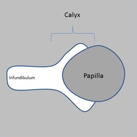
[[File:Lobster-claw-sign-papillary-necrosis-normal.jpg|300px|thumb|left|Normal papillary system.<ref>Case courtesy of Dr Matt A. Morgan, <a href="https://radiopaedia.org/">Radiopaedia.org</a>. From the case <a href="https://radiopaedia.org/cases/40421">rID: 40421</a></ref>]] | [[File:Lobster-claw-sign-papillary-necrosis-normal.jpg|300px|thumb|left|Normal papillary system.<ref>Case courtesy of Dr Matt A. Morgan, <a href="https://radiopaedia.org/">Radiopaedia.org</a>. From the case <a href="https://radiopaedia.org/cases/40421">rID: 40421</a></ref>]] | ||
Revision as of 06:30, 21 August 2020
|
Renal papillary necrosis Microchapters |
|
Differentiating Renal papillary necrosis from other Diseases |
|---|
|
Diagnosis |
|
Treatment |
|
Case Studies |
|
Renal papillary necrosis CT On the Web |
|
American Roentgen Ray Society Images of Renal papillary necrosis CT |
|
Risk calculators and risk factors for Renal papillary necrosis CT |
Please help WikiDoc by adding content here. It's easy! Click here to learn about editing.
Editor-In-Chief: C. Michael Gibson, M.S., M.D. [1]; Associate Editor(s)-in-Chief: Nasrin Nikravangolsefid, MD-MPH [2]
Overview
CT urography is helpful in the diagnosis of renal papillary necrosis that shows multiple small accumulations of contrast media in the papillary regions near the calyx system.
Key CT Findings in Renal papillary necrosis
- CT urography is helpful in the diagnosis of renal papillary necrosis. [1]
- Normal papillary blush is blushlike attenuated area at excretory phase of CT urography. [1]
- Findings on CT urography suggestive of renal papillary necrosis include multiple small accumulations of contrast media in the papillary regions. [1]
- The lobster claw sign is a diagnostic finding on CT urography in the papillary subtype of renal papillary necrosis that develops due to elongation of the minor calyx fornices following ischemia and destruction of the renal papilla. [2]
- Other diagnostic finding include[3]:
- ball on tee
- signet ring
- sloughed papilla with calyces clubbing


References
- ↑ 1.0 1.1 1.2 Kawamoto S, Duggan P, Sheth S, Miyamoto H, Kazi ZN, Fishman EK (2017). "Renal Papillary and Calyceal Lesions at CT Urography: Genitourinary Imaging". Radiographics. 37 (1): 358–359. doi:10.1148/rg.2017160089. PMID 28076022.
- ↑ Xiang H, Han J, Ridley WE, Ridley LJ (2018). "Lobster claw sign: Renal papillary necrosis". J Med Imaging Radiat Oncol. 62 Suppl 1: 90. doi:10.1111/1754-9485.37_12784. PMID 30309154.
- ↑ "Renal papillary necrosis | Radiology Reference Article | Radiopaedia.org".
- ↑ Case courtesy of Dr Matt A. Morgan, <a href="https://radiopaedia.org/">Radiopaedia.org</a>. From the case <a href="https://radiopaedia.org/cases/40421">rID: 40421</a>
- ↑ Case courtesy of Dr Matt A. Morgan, <a href="https://radiopaedia.org/">Radiopaedia.org</a>. From the case <a href="https://radiopaedia.org/cases/40421">rID: 40421</a>