Achalasia CT: Difference between revisions
Jump to navigation
Jump to search
Ahmed Younes (talk | contribs) (→CT) |
Ahmed Younes (talk | contribs) No edit summary |
||
| Line 1: | Line 1: | ||
__NOTOC__ | __NOTOC__ | ||
{{Achalasia}} | {{Achalasia}} | ||
{{CMG}} | {{CMG}}, {{AE}}{{AY}} | ||
==Overview== | ==Overview== | ||
Revision as of 12:47, 3 November 2017
|
Achalasia Microchapters |
|
Diagnosis |
|---|
|
Treatment |
|
Case Studies |
|
Achalasia CT On the Web |
|
American Roentgen Ray Society Images of Achalasia CT |
Editor-In-Chief: C. Michael Gibson, M.S., M.D. [1], Associate Editor(s)-in-Chief: Ahmed Younes M.B.B.CH [2]
Overview
CT
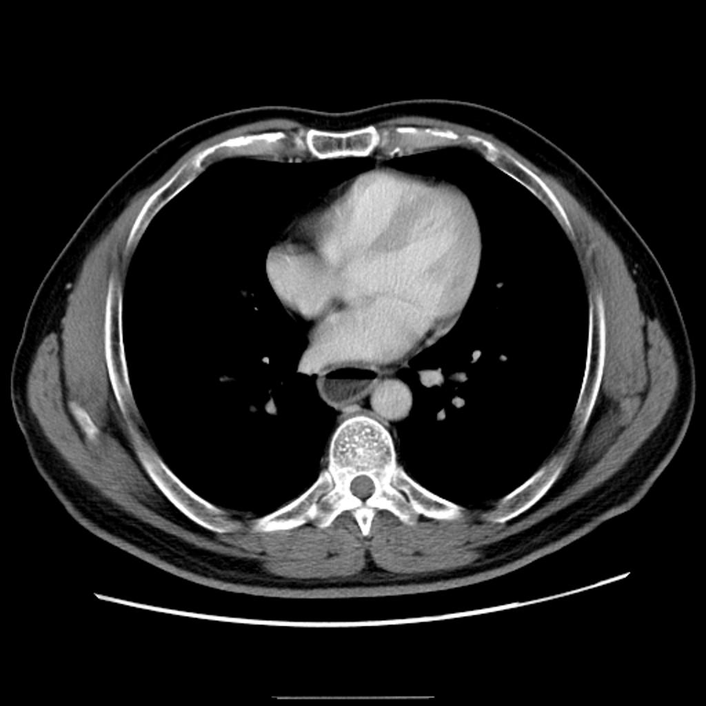
- CT scan may show dilatation of the esophagus with air fluid levels in long-standing cases.
- CT scan may be used to exclude pseudoachalasia, or achalasia symptoms resulting from a different cause, usually esophageal cancer.
