Angiosarcoma: Difference between revisions
Tarek Nafee (talk | contribs) No edit summary |
Tarek Nafee (talk | contribs) No edit summary |
||
| Line 1: | Line 1: | ||
__NOTOC__ | __NOTOC__ | ||
{{SI}} | {{SI}} | ||
{{CMG}}; {{AE}} {{MV | {{CMG}}; {{AE}} {{MV}} | ||
{{SK}} Hemangiosarcoma; Pulmonary angiosarcoma; Vascular sarcoma | {{SK}} Hemangiosarcoma; Pulmonary angiosarcoma; Vascular sarcoma | ||
| Line 10: | Line 10: | ||
==Historical Perspective== | ==Historical Perspective== | ||
Angiosarcoma was first discovered by Dr. Juan Rosai, M.D. and colleagues in 1976.<ref name="pmid24946325">{{cite journal |vauthors=Barber W, Scriven P, Turner D, Hughes D, Wyld D |title=Epithelioid angiosarcoma: Use of angiographic embolisation and radiotherapy to control recurrent haemorrhage |journal=J Surg Case Rep |volume=2010 |issue=5 |pages=7 |year=2010 |pmid=24946325 |pmc=3649120 |doi=10.1093/jscr/2010.5.7 |url=}}</ref> | |||
==Classification== | ==Classification== | ||
Angiosarcoma may be classified according to anatomical location into the following categories: | |||
*Head and neck angiosarcomas | |||
*Skin angiosarcomas (Most common) | |||
*Liver angiosarcomas | |||
*Lung angiosarcomas | |||
*Spleen angiosarcomas | |||
*Others/uncategorized | |||
==Pathophysiology== | ==Pathophysiology== | ||
===Pathogenesis=== | |||
The pathogenesis of angiosarcoma is characterized by a rapid and extensive infiltrating overgrowth of vascular [[epithelial cells]].<ref name="pmid20537949">{{cite journal |vauthors=Young RJ, Brown NJ, Reed MW, Hughes D, Woll PJ |title=Angiosarcoma |journal=Lancet Oncol. |volume=11 |issue=10 |pages=983–91 |year=2010 |pmid=20537949 |doi=10.1016/S1470-2045(10)70023-1 |url=}}</ref> Angiosarcoma is a locally aggressive [[tumor]] with a high rate of [[lymph node]] infiltration and [[metastases]].<ref name="librepathology">Angiosarcoma. Wikipedia. https://en.wikipedia.org/wiki/Angiosarcoma Accessed on April 22, 2016</ref> | |||
===Genetics=== | |||
The [[PTPRB]]/[[Phospholipase C|PLCG1]] [[genes]] are associated with the development of angiosarcoma by triggering aberrant [[angiogenesis]].<ref name="pmid20537949">{{cite journal |vauthors=Young RJ, Brown NJ, Reed MW, Hughes D, Woll PJ |title=Angiosarcoma |journal=Lancet Oncol. |volume=11 |issue=10 |pages=983–91 |year=2010 |pmid=20537949 |doi=10.1016/S1470-2045(10)70023-1 |url=}}</ref> | |||
===Gross Pathology=== | |||
On gross pathology, characteristic findings of angiosarcoma may include:<ref name="pmid20537949">{{cite journal |vauthors=Young RJ, Brown NJ, Reed MW, Hughes D, Woll PJ |title=Angiosarcoma |journal=Lancet Oncol. |volume=11 |issue=10 |pages=983–91 |year=2010 |pmid=20537949 |doi=10.1016/S1470-2045(10)70023-1 |url=}}</ref> | |||
*Red/dark tan lesion | |||
*Typically poorly circumscribed | |||
====Examples of Gross Pathology==== | |||
<gallery> | <gallery> | ||
Image: Angiosarcoma gross pathology.jpg| Gross pathology: angiosarcoma<br> <SMALL> Courtesy of Libre Pathology </SMALL> | Image: Angiosarcoma gross pathology.jpg| Gross pathology: angiosarcoma<br> <SMALL> Courtesy of Libre Pathology </SMALL><ref name="librepathology">Angiosarcoma. Wikipedia. https://en.wikipedia.org/wiki/Angiosarcoma Accessed on April 22, 2016</ref> | ||
</gallery> | </gallery> | ||
===Microscopic Pathology=== | |||
On microscopic histopathological analysis, characteristic findings of angiosarcoma may include:<ref name="librepathology">Angiosarcoma. Wikipedia. https://en.wikipedia.org/wiki/Angiosarcoma Accessed on April 22, 2016</ref> | |||
*[[Spindle cells|Spindle cell]] lesion | |||
*Epitheloid lesion | |||
*Numerous irrergular [[capillaries]] | |||
*Appears red on low power | |||
*[[Pleomorphic]] nuclei | |||
*Hobnail morphology | |||
*Numerous [[mitotic]] bodies | |||
*[[Cytoplasmic]] [[vacuoles]] | |||
*[[Lumen (anatomy)|Luminal]] arrangement of cells | |||
==Causes== | ==Causes== | ||
Common causes of angiosarcoma include:<ref name="librepathology">Angiosarcoma. Wikipedia. https://en.wikipedia.org/wiki/Angiosarcoma Accessed on April 22, 2016</ref><ref name="pmid20537949">{{cite journal |vauthors=Young RJ, Brown NJ, Reed MW, Hughes D, Woll PJ |title=Angiosarcoma |journal=Lancet Oncol. |volume=11 |issue=10 |pages=983–91 |year=2010 |pmid=20537949 |doi=10.1016/S1470-2045(10)70023-1 |url=}}</ref> | |||
*Exposure to [[vinyl chloride]] monomer (VCM) for prolonged periods | |||
*Exposure to [[polyvinyl chloride]] (PVC) polymerisation plants | |||
*Exposure to [[arsenic]]-containing [[insecticides]] | |||
*Previous exposure to [[thorium dioxide]] [[irradiation]] | |||
==Differentiating Angiosarcoma from Other Diseases== | ==Differentiating Angiosarcoma from Other Diseases== | ||
| Line 77: | Line 77: | ||
===Age=== | ===Age=== | ||
*Angiosarcoma is more commonly observed among patients aged between 40 to 75 years old.<ref name="angio">Sturgis EM, Potter BO. Sarcomas of the head and neck region. Curr Opin Oncol. 2003 May. 15(3):239-52</ref> | *Angiosarcoma is more commonly observed among patients aged between 40 to 75 years old.<ref name="angio">Sturgis EM, Potter BO. Sarcomas of the head and neck region. Curr Opin Oncol. 2003 May. 15(3):239-52</ref> | ||
===Gender=== | ===Gender=== | ||
| Line 85: | Line 83: | ||
===Race=== | ===Race=== | ||
There is no racial predilection for angiosarcoma. | |||
==Risk Factors== | ==Risk Factors== | ||
Common risk factors in the development of angiosarcoma include:<ref name="pmid20537949">{{cite journal |vauthors=Young RJ, Brown NJ, Reed MW, Hughes D, Woll PJ |title=Angiosarcoma |journal=Lancet Oncol. |volume=11 |issue=10 |pages=983–91 |year=2010 |pmid=20537949 |doi=10.1016/S1470-2045(10)70023-1 |url=}}</ref> | |||
*Chronic lymphedema | |||
*Chronic exposure to [[polyvinyl chloride]] (PVC) | |||
*[[Radiation exposure]] | |||
*Exposure to [[Thorotrast]] | |||
== Natural History, Complications and Prognosis== | == Natural History, Complications and Prognosis== | ||
===Natural History=== | |||
The majority of patients with angiosarcoma remain asymptomatic for years.<ref name="pmid20537949">{{cite journal |vauthors=Young RJ, Brown NJ, Reed MW, Hughes D, Woll PJ |title=Angiosarcoma |journal=Lancet Oncol. |volume=11 |issue=10 |pages=983–91 |year=2010 |pmid=20537949 |doi=10.1016/S1470-2045(10)70023-1 |url=}}</ref> Early clinical features may include nonspecific symptoms, such as [[pain]], [[fatigue]], [[malaise]], and [[nausea]]. If left untreated, the majority of patients with angiosarcoma may rapidly progress to develop [[metastases]].<ref name="angio">Sturgis EM, Potter BO. Sarcomas of the head and neck region. Curr Opin Oncol. 2003 May. 15(3):239-52</ref> | |||
===Complications=== | |||
Common complications of angiosarcoma include:<ref name="pmid20537949">{{cite journal |vauthors=Young RJ, Brown NJ, Reed MW, Hughes D, Woll PJ |title=Angiosarcoma |journal=Lancet Oncol. |volume=11 |issue=10 |pages=983–91 |year=2010 |pmid=20537949 |doi=10.1016/S1470-2045(10)70023-1 |url=}}</ref> | |||
*[[Fractures|Pathologic fractures]] | |||
*[[Anemia]] | |||
*[[Hepatic dysfunction]] | |||
===Prognosis=== | |||
*Poor prognostic factors include patient age (> 65 years), [[retroperitoneal]] location, and large [[tumor]] size.<ref name="angio">Sturgis EM, Potter BO. Sarcomas of the head and neck region. Curr Opin Oncol. 2003 May. 15(3):239-52</ref> | Prognosis is generally poor; the 5-year survival rate of patients with angiosarcoma is approximately 12-33%. *Poor prognostic factors include patient age (> 65 years), [[retroperitoneal]] location, and large [[tumor]] size.<ref name="angio">Sturgis EM, Potter BO. Sarcomas of the head and neck region. Curr Opin Oncol. 2003 May. 15(3):239-52</ref> | ||
== Diagnosis == | == Diagnosis == | ||
=== Symptoms === | === Symptoms === | ||
Angiosarcoma is usually asymptomatic and found incidentally. Symptoms of angiosarcoma are generally non-specific. | |||
=== Physical Examination === | === Physical Examination === | ||
Patients with angiosarcoma may appear [[Cachexia|cachectic]] or normal. In cutaneous angiosarcoma, physical examination findings may include: | |||
*Bruise or [[skin ulceration]] | |||
*Blushed purple-red [[papule]] | |||
=== Laboratory Findings === | === Laboratory Findings === | ||
There are no specific laboratory findings associated with angiosarcoma. | |||
===Imaging Findings=== | ===Imaging Findings=== | ||
The imaging modality of choice for angiosarcoma will depend on the location. For pulmonary angiosarcoma, the imaging modality of choice is enhanced CT scan.<ref name="angio">Sturgis EM, Potter BO. Sarcomas of the head and neck region. Curr Opin Oncol. 2003 May. 15(3):239-52</ref> For other types angiosarcoma, the imaging modality of choice is MRI. | |||
====CT==== | ====CT==== | ||
On CT, findings of angiosarcoma may include:<ref name="angio">Sturgis EM, Potter BO. Sarcomas of the head and neck region. Curr Opin Oncol. 2003 May. 15(3):239-52</ref> | On CT, findings of angiosarcoma may include:<ref name="angio">Sturgis EM, Potter BO. Sarcomas of the head and neck region. Curr Opin Oncol. 2003 May. 15(3):239-52</ref> | ||
| Line 136: | Line 128: | ||
====MRI==== | ====MRI==== | ||
On MRI, findings of angiosarcoma may include: | On MRI, findings of angiosarcoma may include: | ||
*T1/T2: heterogeneous areas of hyperintensity suggestive of a mixed tumour and [[hemorrhage]] | |||
*T1 C+ (Gd): heterogeneous enhancement | |||
== Treatment == | == Treatment == | ||
=== Medical Therapy === | === Medical Therapy === | ||
The main [[adjuvant therapy]] for angiosarcoma is a [[doxorubicin]]-based regimen.<ref name="pmid20537949">{{cite journal |vauthors=Young RJ, Brown NJ, Reed MW, Hughes D, Woll PJ |title=Angiosarcoma |journal=Lancet Oncol. |volume=11 |issue=10 |pages=983–91 |year=2010 |pmid=20537949 |doi=10.1016/S1470-2045(10)70023-1 |url=}}</ref> For angiosarcoma, [[doxorubicin]] monotherapy is indicated as first-line therapy.<ref name="pmid20537949">{{cite journal |vauthors=Young RJ, Brown NJ, Reed MW, Hughes D, Woll PJ |title=Angiosarcoma |journal=Lancet Oncol. |volume=11 |issue=10 |pages=983–91 |year=2010 |pmid=20537949 |doi=10.1016/S1470-2045(10)70023-1 |url=}}</ref> | |||
*Common complications of [[doxorubicin]] include: | |||
:*[[Cardiotoxicity]] | |||
:*[[Mucositis]] | |||
:*[[Alopecia]] | |||
:*[[Nausea]] | |||
:*[[Vomiting]] | |||
For patients with pulmonary angiosarcoma, a combination of [[radiotherapy]] and [[cancer immunotherapy|immunotherapy]] with recombinant [[interleukin-2]] is the treatment of choice.<ref name="pmid15249484">{{cite journal |vauthors=Duck L, Baurain JF, Machiels JP |title=Treatment of a primary pulmonary angiosarcoma |journal=Chest |volume=126 |issue=1 |pages=317–8; author reply 318 |year=2004 |pmid=15249484 |doi=10.1378/chest.126.1.317 |url=}}</ref> The response rate to [[chemotherapy]] in patients with angiosarcoma is poor.<ref name="pmid20537949">{{cite journal |vauthors=Young RJ, Brown NJ, Reed MW, Hughes D, Woll PJ |title=Angiosarcoma |journal=Lancet Oncol. |volume=11 |issue=10 |pages=983–91 |year=2010 |pmid=20537949 |doi=10.1016/S1470-2045(10)70023-1 |url=}}</ref> | |||
=== Surgery === | === Surgery === | ||
Surgical resection in combination with [[radiation therapy]] is the treatment of choice for small angiosarcomas.<ref name="pmid20537949">{{cite journal |vauthors=Young RJ, Brown NJ, Reed MW, Hughes D, Woll PJ |title=Angiosarcoma |journal=Lancet Oncol. |volume=11 |issue=10 |pages=983–91 |year=2010 |pmid=20537949 |doi=10.1016/S1470-2045(10)70023-1 |url=}}</ref> | |||
*Surgical treatment for patients with cutaneous angiosarcoma is surgical resection with wide margins.<ref name="pmid20537949">{{cite journal |vauthors=Young RJ, Brown NJ, Reed MW, Hughes D, Woll PJ |title=Angiosarcoma |journal=Lancet Oncol. |volume=11 |issue=10 |pages=983–91 |year=2010 |pmid=20537949 |doi=10.1016/S1470-2045(10)70023-1 |url=}}</ref> | *Surgical treatment for patients with cutaneous angiosarcoma is surgical resection with wide margins.<ref name="pmid20537949">{{cite journal |vauthors=Young RJ, Brown NJ, Reed MW, Hughes D, Woll PJ |title=Angiosarcoma |journal=Lancet Oncol. |volume=11 |issue=10 |pages=983–91 |year=2010 |pmid=20537949 |doi=10.1016/S1470-2045(10)70023-1 |url=}}</ref> Surgery is not recommended on patients with large sized angiosarcomas. The recurrence rate of angiosarcoma after surgery is 80%. | ||
=== Prevention === | === Prevention === | ||
====Primary Prevention==== | |||
There are no primary preventive measures available for angiosarcoma. | |||
====Secondary Prevention==== | |||
Once diagnosed and successfully treated, patients with angiosarcoma are followed-up every 3, 6, or 12 months depending on the stage at diagnosis. Follow-up testing for angiosarcoma may include:<ref name="pmid20537949">{{cite journal |vauthors=Young RJ, Brown NJ, Reed MW, Hughes D, Woll PJ |title=Angiosarcoma |journal=Lancet Oncol. |volume=11 |issue=10 |pages=983–91 |year=2010 |pmid=20537949 |doi=10.1016/S1470-2045(10)70023-1 |url=}}</ref> | |||
*Periodic imaging/[[angiography]] evaluation | |||
*Laboratory testing: [[complete blood count]] (e.g., [[anemia]]) | |||
==References== | ==References== | ||
| Line 171: | Line 160: | ||
[[Category:Oncology]] | [[Category:Oncology]] | ||
[[Category:FinalQCRequired]] | [[Category:FinalQCRequired]] | ||
{{WH}}{{WS}} | |||
Revision as of 19:00, 24 August 2016
|
WikiDoc Resources for Angiosarcoma |
|
Articles |
|---|
|
Most recent articles on Angiosarcoma Most cited articles on Angiosarcoma |
|
Media |
|
Powerpoint slides on Angiosarcoma |
|
Evidence Based Medicine |
|
Clinical Trials |
|
Ongoing Trials on Angiosarcoma at Clinical Trials.gov Clinical Trials on Angiosarcoma at Google
|
|
Guidelines / Policies / Govt |
|
US National Guidelines Clearinghouse on Angiosarcoma
|
|
Books |
|
News |
|
Commentary |
|
Definitions |
|
Patient Resources / Community |
|
Patient resources on Angiosarcoma Discussion groups on Angiosarcoma Patient Handouts on Angiosarcoma Directions to Hospitals Treating Angiosarcoma Risk calculators and risk factors for Angiosarcoma
|
|
Healthcare Provider Resources |
|
Causes & Risk Factors for Angiosarcoma |
|
Continuing Medical Education (CME) |
|
International |
|
|
|
Business |
|
Experimental / Informatics |
Editor-In-Chief: C. Michael Gibson, M.S., M.D. [1]; Associate Editor(s)-in-Chief: Maria Fernanda Villarreal, M.D. [2]
Synonyms and keywords: Hemangiosarcoma; Pulmonary angiosarcoma; Vascular sarcoma
Overview
Angiosarcoma is a rare malignant vascular neoplasm of endothelial-type cells that line vessel walls.[1][2] Angiosarcoma was first discovered by Dr. Juan Rosai, M.D. and colleagues in 1976.[3] The pathogenesis of angiosarcoma is characterized by a rapid and extensively infiltrating overgrowth of vascular epithelial cells. Common angiosarcoma locations include the kidney, liver, lung, breast, and liver. The PTPRB/PLCG1 genes are associated with the development of angiosarcoma; the mutation results in aberrant angiogenesis. The imaging modality of choice for diagnosing angiosarcoma will depend on the location. For pulmonary angiosarcoma, the imaging modality of choice is enhanced CT scan.[4] For other types angiosarcoma, the imaging modality of choice is MRI. On CT scan, findings suggestive of angiosarcoma may include vascular invasion, nodular enhancement (common), and a hypoattenuating mass. The mainstay adjuvant therapy for angiosarcoma is a doxorubicin-based regimen.[5] The response rate for chemotherapy in patients with angiosarcoma is poor.[5]
Historical Perspective
Angiosarcoma was first discovered by Dr. Juan Rosai, M.D. and colleagues in 1976.[3]
Classification
Angiosarcoma may be classified according to anatomical location into the following categories:
- Head and neck angiosarcomas
- Skin angiosarcomas (Most common)
- Liver angiosarcomas
- Lung angiosarcomas
- Spleen angiosarcomas
- Others/uncategorized
Pathophysiology
Pathogenesis
The pathogenesis of angiosarcoma is characterized by a rapid and extensive infiltrating overgrowth of vascular epithelial cells.[5] Angiosarcoma is a locally aggressive tumor with a high rate of lymph node infiltration and metastases.[2]
Genetics
The PTPRB/PLCG1 genes are associated with the development of angiosarcoma by triggering aberrant angiogenesis.[5]
Gross Pathology
On gross pathology, characteristic findings of angiosarcoma may include:[5]
- Red/dark tan lesion
- Typically poorly circumscribed
Examples of Gross Pathology
-
![Gross pathology: angiosarcoma Courtesy of Libre Pathology [2]](/images/2/21/Angiosarcoma_gross_pathology.jpg)
Gross pathology: angiosarcoma
Courtesy of Libre Pathology [2]
![Gross pathology: angiosarcoma Courtesy of Libre Pathology [2]](/index.php/File:Angiosarcoma_gross_pathology.jpg)
Microscopic Pathology
On microscopic histopathological analysis, characteristic findings of angiosarcoma may include:[2]
- Spindle cell lesion
- Epitheloid lesion
- Numerous irrergular capillaries
- Appears red on low power
- Pleomorphic nuclei
- Hobnail morphology
- Numerous mitotic bodies
- Cytoplasmic vacuoles
- Luminal arrangement of cells
Causes
Common causes of angiosarcoma include:[2][5]
- Exposure to vinyl chloride monomer (VCM) for prolonged periods
- Exposure to polyvinyl chloride (PVC) polymerisation plants
- Exposure to arsenic-containing insecticides
- Previous exposure to thorium dioxide irradiation
Differentiating Angiosarcoma from Other Diseases
Angiosarcoma must be differentiated from other diseases that cause a highly vascular mass or non-healing cutaneous ulcerations such as:[6]
Differentials for Cutaneous Angiosarcoma
Cutaneous angiosarcoma must be differentiated from other diseases with non-healing cutaneous ulcerations such as:
Differentials for Non-cutaneous Angiosarcoma
Angiosarcoma must be differentiated from other diseases that cause a highly vascular mass such as:
- Atypical vascular lesions
- Hemangioma
- Glomangiosarcoma
- Carotid body tumor
- Malignant fibrous histiocytoma of soft tissue
Epidemiology and Demographics
Incidence
In 2004, the age-adjusted incidence of angiosarcoma was 3.1 per 100,000 population per year.[4]
Age
- Angiosarcoma is more commonly observed among patients aged between 40 to 75 years old.[4]
Gender
- Males are more commonly affected with angiosarcoma than females.[4]
- The male to female ratio is 2:1.[4]
Race
There is no racial predilection for angiosarcoma.
Risk Factors
Common risk factors in the development of angiosarcoma include:[5]
- Chronic lymphedema
- Chronic exposure to polyvinyl chloride (PVC)
- Radiation exposure
- Exposure to Thorotrast
Natural History, Complications and Prognosis
Natural History
The majority of patients with angiosarcoma remain asymptomatic for years.[5] Early clinical features may include nonspecific symptoms, such as pain, fatigue, malaise, and nausea. If left untreated, the majority of patients with angiosarcoma may rapidly progress to develop metastases.[4]
Complications
Common complications of angiosarcoma include:[5]
Prognosis
Prognosis is generally poor; the 5-year survival rate of patients with angiosarcoma is approximately 12-33%. *Poor prognostic factors include patient age (> 65 years), retroperitoneal location, and large tumor size.[4]
Diagnosis
Symptoms
Angiosarcoma is usually asymptomatic and found incidentally. Symptoms of angiosarcoma are generally non-specific.
Physical Examination
Patients with angiosarcoma may appear cachectic or normal. In cutaneous angiosarcoma, physical examination findings may include:
- Bruise or skin ulceration
- Blushed purple-red papule
Laboratory Findings
There are no specific laboratory findings associated with angiosarcoma.
Imaging Findings
The imaging modality of choice for angiosarcoma will depend on the location. For pulmonary angiosarcoma, the imaging modality of choice is enhanced CT scan.[4] For other types angiosarcoma, the imaging modality of choice is MRI.
CT
On CT, findings of angiosarcoma may include:[4]
- Vascular invasion
- Nodular enhancement (common)
- Hypoattenuating mass
- Multicentric lesions
-
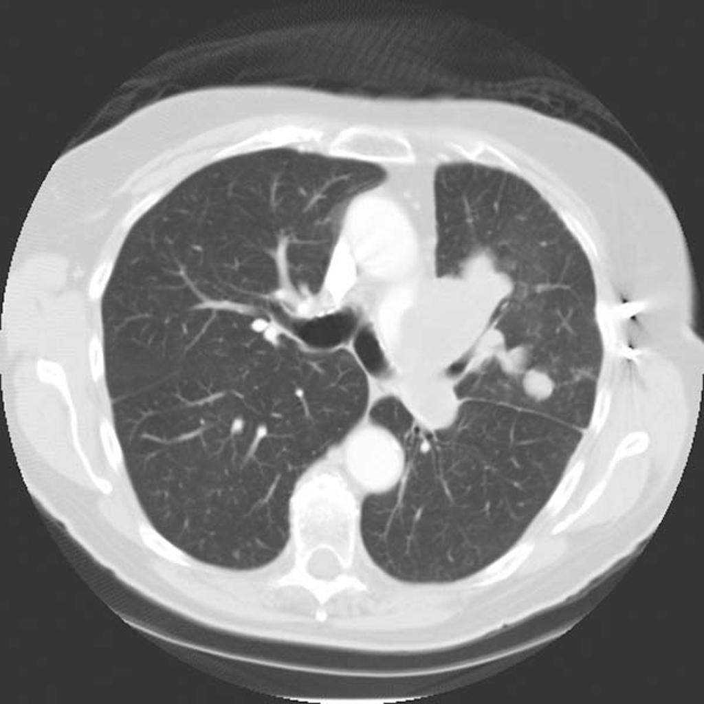
CT Pulmonary angiosarcoma
Courtesy of Radiopedia

MRI
On MRI, findings of angiosarcoma may include:
- T1/T2: heterogeneous areas of hyperintensity suggestive of a mixed tumour and hemorrhage
- T1 C+ (Gd): heterogeneous enhancement
Treatment
Medical Therapy
The main adjuvant therapy for angiosarcoma is a doxorubicin-based regimen.[5] For angiosarcoma, doxorubicin monotherapy is indicated as first-line therapy.[5]
- Common complications of doxorubicin include:
For patients with pulmonary angiosarcoma, a combination of radiotherapy and immunotherapy with recombinant interleukin-2 is the treatment of choice.[7] The response rate to chemotherapy in patients with angiosarcoma is poor.[5]
Surgery
Surgical resection in combination with radiation therapy is the treatment of choice for small angiosarcomas.[5]
- Surgical treatment for patients with cutaneous angiosarcoma is surgical resection with wide margins.[5] Surgery is not recommended on patients with large sized angiosarcomas. The recurrence rate of angiosarcoma after surgery is 80%.
Prevention
Primary Prevention
There are no primary preventive measures available for angiosarcoma.
Secondary Prevention
Once diagnosed and successfully treated, patients with angiosarcoma are followed-up every 3, 6, or 12 months depending on the stage at diagnosis. Follow-up testing for angiosarcoma may include:[5]
- Periodic imaging/angiography evaluation
- Laboratory testing: complete blood count (e.g., anemia)
References
- ↑ Perkins, [edited by] Vinay Kumar, Abul K. Abbas, Jon C. Aster ; artist, James A. (2013). Robbins basic pathology (9th ed. ed.). Philadelphia, PA: Elsevier/Saunders. ISBN 9781437717815.
- ↑ 2.0 2.1 2.2 2.3 2.4 Angiosarcoma. Wikipedia. https://en.wikipedia.org/wiki/Angiosarcoma Accessed on April 22, 2016
- ↑ 3.0 3.1 Barber W, Scriven P, Turner D, Hughes D, Wyld D (2010). "Epithelioid angiosarcoma: Use of angiographic embolisation and radiotherapy to control recurrent haemorrhage". J Surg Case Rep. 2010 (5): 7. doi:10.1093/jscr/2010.5.7. PMC 3649120. PMID 24946325.
- ↑ 4.0 4.1 4.2 4.3 4.4 4.5 4.6 4.7 4.8 Sturgis EM, Potter BO. Sarcomas of the head and neck region. Curr Opin Oncol. 2003 May. 15(3):239-52
- ↑ 5.00 5.01 5.02 5.03 5.04 5.05 5.06 5.07 5.08 5.09 5.10 5.11 5.12 5.13 5.14 Young RJ, Brown NJ, Reed MW, Hughes D, Woll PJ (2010). "Angiosarcoma". Lancet Oncol. 11 (10): 983–91. doi:10.1016/S1470-2045(10)70023-1. PMID 20537949.
- ↑ Angiosarcoma. Wikipedia. https://en.wikipedia.org/wiki/Angiosarcoma Accessed April 22, 2016
- ↑ Duck L, Baurain JF, Machiels JP (2004). "Treatment of a primary pulmonary angiosarcoma". Chest. 126 (1): 317–8, author reply 318. doi:10.1378/chest.126.1.317. PMID 15249484.