Sandbox: Singlepage Maria: Difference between revisions
No edit summary |
|||
| Line 7: | Line 7: | ||
==Overview== | ==Overview== | ||
'''Ductal carcinoma''' is the most common type of breast cancer in women. Ductal carcinoma may be classified according to the Armed Forces Institute of Pathology (AFIP) into | '''Ductal carcinoma''' is the most common type of breast cancer in women. Ductal carcinoma may be classified according to the Armed Forces Institute of Pathology (AFIP) into 2 groups: large cell carcinoma in situ and small cell carcinoma in situ. The pathogenesis of ductal carcinoma is characterized by the microinvasion of cancer cells limited to the ducts with no extension beyond the basement membrane. The mutation on HER2/neu has been associated with the development of ductal carcinoma. | ||
==Historical Perspective== | ==Historical Perspective== | ||
| Line 14: | Line 13: | ||
==Classification== | ==Classification== | ||
*Ductal carcinoma may be classified according to the Armed Forces Institute of Pathology (AFIP) into | *Ductal carcinoma may be classified according to the Armed Forces Institute of Pathology (AFIP) into 2 groups: | ||
:* | |||
:* | :*'''Large cell''' | ||
:* | ::*More aggressive form | ||
::*Also referred to as comedocarcinoma | |||
:*'''Small cell''' | |||
::*Less aggressive | |||
::*Subtypes include cribriform, micropapillary, papillary, and solid in situ. | |||
*Other variants of ductal carcinoma include, non-DCIS entities. | *Other variants of ductal carcinoma include, non-DCIS entities. | ||
==Pathophysiology== | ==Pathophysiology== | ||
*The pathogenesis of ductal carcinoma is characterized by the microinvasion of cancer cells to the ducts | *The pathogenesis of ductal carcinoma is characterized by the microinvasion of cancer cells limited to the ducts with no extension beyond the basement membrane. | ||
*The mutation on HER2/neu has been associated with the development of ductal carcinoma. | *The mutation on HER2/neu has been associated with the development of ductal carcinoma. | ||
*On gross pathology, characteristic findings of ductal carcinoma, include: | *On gross pathology, characteristic findings of ductal carcinoma, include: | ||
| Line 105: | Line 109: | ||
=== Laboratory Findings === | === Laboratory Findings === | ||
*Laboratory findings consistent with the diagnosis of ductal carcinoma, include: | *Laboratory findings consistent with the diagnosis of ductal carcinoma, include: | ||
:*Positive/negative estrogen receptor (ER) and progesterone receptor (PR) expression | |||
===Imaging Findings=== | ===Imaging Findings=== | ||
*Mammography is the imaging modality of choice for ductal carcinoma. | *Mammography is the imaging modality of choice for ductal carcinoma. | ||
*On mammography, findings of ductal carcinoma, include: | *On mammography, findings of ductal carcinoma, include: | ||
:*Calcifications (most common) | |||
:*Simple mass | |||
:*Soft-tissue opacity | |||
:*Asymmetry without calcification | |||
*The image below demonstrates findings compatible with ductal carcinoma. | |||
<gallery> | |||
Image:Mammo breast cancer.jpg|Normal (left) versus cancerous (right) mammography image. | |||
</gallery> | |||
=== Other Diagnostic Studies === | === Other Diagnostic Studies === | ||
Revision as of 15:54, 19 April 2016
Editor-In-Chief: C. Michael Gibson, M.S., M.D. [1] Associate Editor(s)-in-Chief: Maria Fernanda Villarreal, M.D. [2]
Synonyms and keywords: Intraductal hyperplasia; IDH; Atypical ductal hyperplasia; Comedocarcinoma; Duct cell carcinoma; Duct carcinoma
Overview
Ductal carcinoma is the most common type of breast cancer in women. Ductal carcinoma may be classified according to the Armed Forces Institute of Pathology (AFIP) into 2 groups: large cell carcinoma in situ and small cell carcinoma in situ. The pathogenesis of ductal carcinoma is characterized by the microinvasion of cancer cells limited to the ducts with no extension beyond the basement membrane. The mutation on HER2/neu has been associated with the development of ductal carcinoma.
Historical Perspective
Ductal carcinoma was first described in 1893.
Classification
- Ductal carcinoma may be classified according to the Armed Forces Institute of Pathology (AFIP) into 2 groups:
- Large cell
- More aggressive form
- Also referred to as comedocarcinoma
- Small cell
- Less aggressive
- Subtypes include cribriform, micropapillary, papillary, and solid in situ.
- Other variants of ductal carcinoma include, non-DCIS entities.
Pathophysiology
- The pathogenesis of ductal carcinoma is characterized by the microinvasion of cancer cells limited to the ducts with no extension beyond the basement membrane.
- The mutation on HER2/neu has been associated with the development of ductal carcinoma.
- On gross pathology, characteristic findings of ductal carcinoma, include:
- White
- Firm stellate lesion
- On microscopic histopathological analysis, characteristic findings of ductal carcinoma, include:
- Equal spacing of cells - "cookie cutter" look.
- Cells line-up along lumen/glandular spaces - form "Roman briges".
- Nuclear enlargement (key feature)
Causes
- Common causes of ductal carcinoma, may include:
Differentiating ductal carcinoma from other Diseases
- Ductal carcinoma must be differentiated from other diseases that cause nipple discharge, breast skin color change, and palpable mass such as:
- [Differential dx1]
- [Differential dx2]
- [Differential dx3]
Epidemiology and Demographics
- The prevalence of ductal carcinoma is approximately 32.5 per 100,000 women worldwide.
- In [year], the incidence of ductal carcinoma was estimated to be [number or range] cases per 100,000 individuals in [location].
Age
- Ductal carcinoma is commonly observed among females between 40 to 80 years old
- Ductal carcinoma is rarely observed among males between 60 and 70 years of age
- Ductal carcinoma is more commonly observed among postmenopausal women
Gender
- Ductal carcinoma affects men and women equally.
- [Gender 1] are more commonly affected with ductal carcinoma than [gender 2].
- The [gender 1] to [Gender 2] ratio is approximately [number > 1] to 1.
Race
- There is no racial predilection for ductal carcinoma.
Risk Factors
- Common risk factors in the development of ductal carcinoma, include:
- Family history of breast cancer
- Mutations in BRCA1/BRCA2 genes
- Previous exposure to radiation therapy
- Increased breast density
- Hormonal therapy
- Nulliparity
- Obesity
Natural History, Complications and Prognosis
- The majority of patients with ductal carcinoma remain asymptomatic for years.
- Early clinical features include skin color change or nipple discharge.
- If left untreated, the majority of patients with ductal carcinoma may progress to develop lymph node invasion, and metastasis.
- The most common complication of ductal carcinoma is lymphedema.
- Prognosis generally depends on the histological subtype.
- In general, the 20-year mortality rate among patients with ductal carcinoma is approximately 3.3%.
- Factors related with worse prognosis, include: young age at diagnosis, black ethnicity, and high grade cancer.
Diagnosis
Diagnostic Criteria
- The diagnosis of ductal carcinoma is made when at least [number] of the following [number] diagnostic criteria are met:
- [criterion 1]
- [criterion 2]
- [criterion 3]
- [criterion 4]
Symptoms
- Ductal carcinoma is usually asymptomatic.
- Symptoms of ductal carcinoma may include the following:
- Nipple discharge
- Skin color changes
- Warm and thickened
- Skin of an orange appearance
- Nipple retraction
Physical Examination
- Patients with ductal carcinoma usually are well-appearing.
- Physical examination may show no specific physical findings.
- In some cases, it may be remarkable for:
- Palpable mass
Laboratory Findings
- Laboratory findings consistent with the diagnosis of ductal carcinoma, include:
- Positive/negative estrogen receptor (ER) and progesterone receptor (PR) expression
Imaging Findings
- Mammography is the imaging modality of choice for ductal carcinoma.
- On mammography, findings of ductal carcinoma, include:
- Calcifications (most common)
- Simple mass
- Soft-tissue opacity
- Asymmetry without calcification
- The image below demonstrates findings compatible with ductal carcinoma.
-
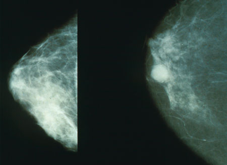
Normal (left) versus cancerous (right) mammography image.

Other Diagnostic Studies
- Ductal carcinoma may also be diagnosed using [diagnostic study name].
- Findings on [diagnostic study name] include [finding 1], [finding 2], and [finding 3].
Treatment
Medical Therapy
- The mainstay of therapies for ductal carcinoma are divided into 2 groups: hormonal therapy and targeted therapy.
Hormonal Therapy
- Selective estrogen receptor modulators, such as:
- Tamoxifen
- Raloxifene
Targeted Therapy
- HER2-directed therapy
- Trastuzumab
- The primary goal of medical therapy is to reduce the risk of ipsilateral or contralateral breast invasion and also decreases the risk of recurrence.
- Response to medical therapy can be monitored with [test/physical finding/imaging] every [frequency/duration].
Surgery
- Surgery is the mainstay of therapy for ductal carcinoma.
- Surgical approaches for ductal carcinoma, include: mastectomy or breast-conserving therapy
- [Surgical procedure] in conjunction with [chemotherapy/radiation] is the most common approach to the treatment of ductal carcinoma.
- [Surgical procedure] can only be performed for patients with [disease stage] ductal carcinoma.
Prevention
- Effective measures for the secondary prevention of ductal carcinoma include: screening mammography for women between 50-74 years and periodical breast self-examination (BSE). [1]
- Once diagnosed and successfully treated, patients with ductal carcinoma are followed-up every 3, 6, or 12 months depending on individual assessment.
- Follow-up testing includes [test 1], [test 2], and [test 3].
References
- ↑ US Task Preventive Force. http://www.uspreventiveservicestaskforce.org/Page/Document/RecommendationStatementFinal/breast-cancer-screening Accessed on April 19, 2016