Osteochondroma CT: Difference between revisions
Jump to navigation
Jump to search
No edit summary |
No edit summary |
||
| Line 13: | Line 13: | ||
==Gallery== | ==Gallery== | ||
<div align="left"> | |||
<gallery heights="175" widths="175"> | |||
Image: Osteochondroma-1(1).jpg|Volume rendered CT : femur osteochondroma | |||
Image: Osteochondroma-femur(2).jpg|CT (coronal view): femur osteochondroma | |||
</gallery> | |||
</div> | |||
Revision as of 16:41, 29 January 2016
|
Osteochondroma Microchapters |
|
Diagnosis |
|---|
|
Treatment |
|
Case Studies |
|
Osteochondroma CT On the Web |
|
American Roentgen Ray Society Images of Osteochondroma CT |
Editor-In-Chief: C. Michael Gibson, M.S., M.D. [1]Associate Editor(s)-in-Chief: Maria Fernanda Villarreal, M.D. [2]
Overview
On CT scan, osteochondroma shows the same findings as on radiograph, but it has better accuracy to demonstrate medullary continuity and the cartilage cap.[1]
CT
CT findings associated with osteochondroma, include:[1]
- Visualization of the mineralization in the cartilage cap
- Evaluation of the marrow continuity of the lesion
Gallery
-
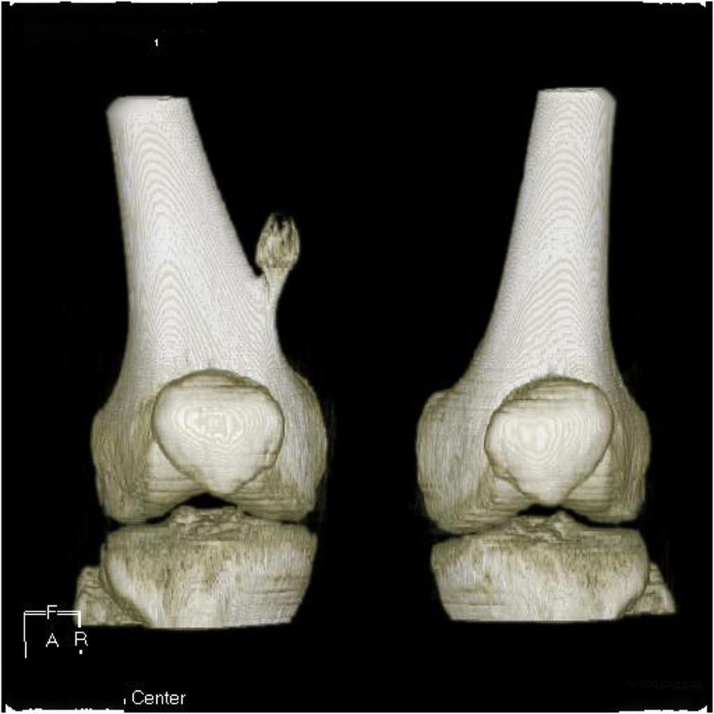
Volume rendered CT : femur osteochondroma
-
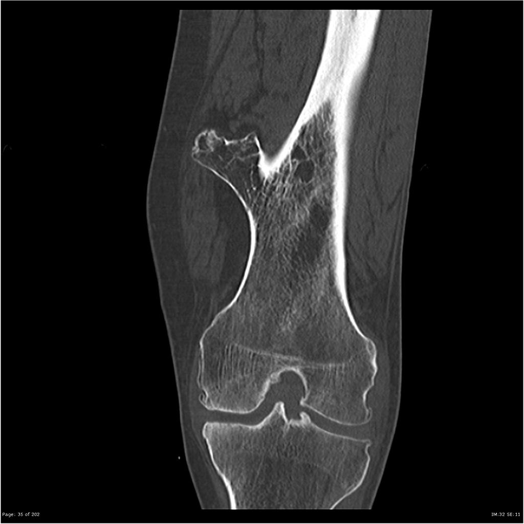
CT (coronal view): femur osteochondroma
.jpg)
.jpg)
References
- ↑ 1.0 1.1 Osteochondroma. Dr Yuranga Weerakkody. Radiopedia. http://radiopaedia.org/articles/osteochondroma Accessed on January 28, 2016