Mycosis fungoides pathophysiology: Difference between revisions
No edit summary |
No edit summary |
||
| Line 3: | Line 3: | ||
{{CMG}}; {{AE}} {{AS}} | {{CMG}}; {{AE}} {{AS}} | ||
==Overview== | ==Overview== | ||
Cutaneous T cell lymphoma arises from [[T-cells]]. On microscopic histopathological analysis, atypical [[lymphoid]] cells, [[polymorphous]] inflammatory infiltrate in the dermis, and [[lymphocytes]] with cerebroid nuclei are characteristic findings of mycosis fungoides. | Cutaneous T cell lymphoma arises from [[T-cells]] lymphocytes. On microscopic histopathological analysis, atypical [[lymphoid]] cells, [[polymorphous]] inflammatory infiltrate in the dermis, and [[lymphocytes]] with cerebroid nuclei are characteristic findings of mycosis fungoides. | ||
==Pathophysiology== | ==Pathophysiology== | ||
* Cutaneous T cell lymphoma is an unusual expression of [[T-cells]], a part of the immune system | * Cutaneous T cell lymphoma is an unusual expression of [[T-cells]], a part of the immune system | ||
Revision as of 16:31, 27 January 2016
|
Cutaneous T cell lymphoma Microchapters |
Editor-In-Chief: C. Michael Gibson, M.S., M.D. [1]; Associate Editor(s)-in-Chief: Sowminya Arikapudi, M.B,B.S. [2]
Overview
Cutaneous T cell lymphoma arises from T-cells lymphocytes. On microscopic histopathological analysis, atypical lymphoid cells, polymorphous inflammatory infiltrate in the dermis, and lymphocytes with cerebroid nuclei are characteristic findings of mycosis fungoides.
Pathophysiology
- Cutaneous T cell lymphoma is an unusual expression of T-cells, a part of the immune system
- These T-cells are skin-associated, meaning that they biochemically and biologically are most related to the skin, in a dynamic manner
- Sezary syndrome and mycosis fungoides are T-cell lymphomas whose primary manifestation is in the skin
- Mycosis Fungoides is the most common type of 'cutaneous T cell lymphoma' (CTCL)
- Mycosis fungoides is initially an indolent lymphoma but in its later stages can cause peripheral lymphadenopathy and can finally progress to widespread extracutaneous visceral / internal organ involvement
- Sézary's cells are T-cells that have pathological quantities of mucopolysaccharides
- Sézary's disease is sometimes considered a late stage of mycosis fungoides
Gross Pathology
-
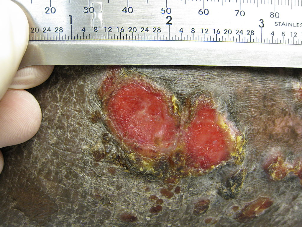
Plaque of mycosis fungoides
-
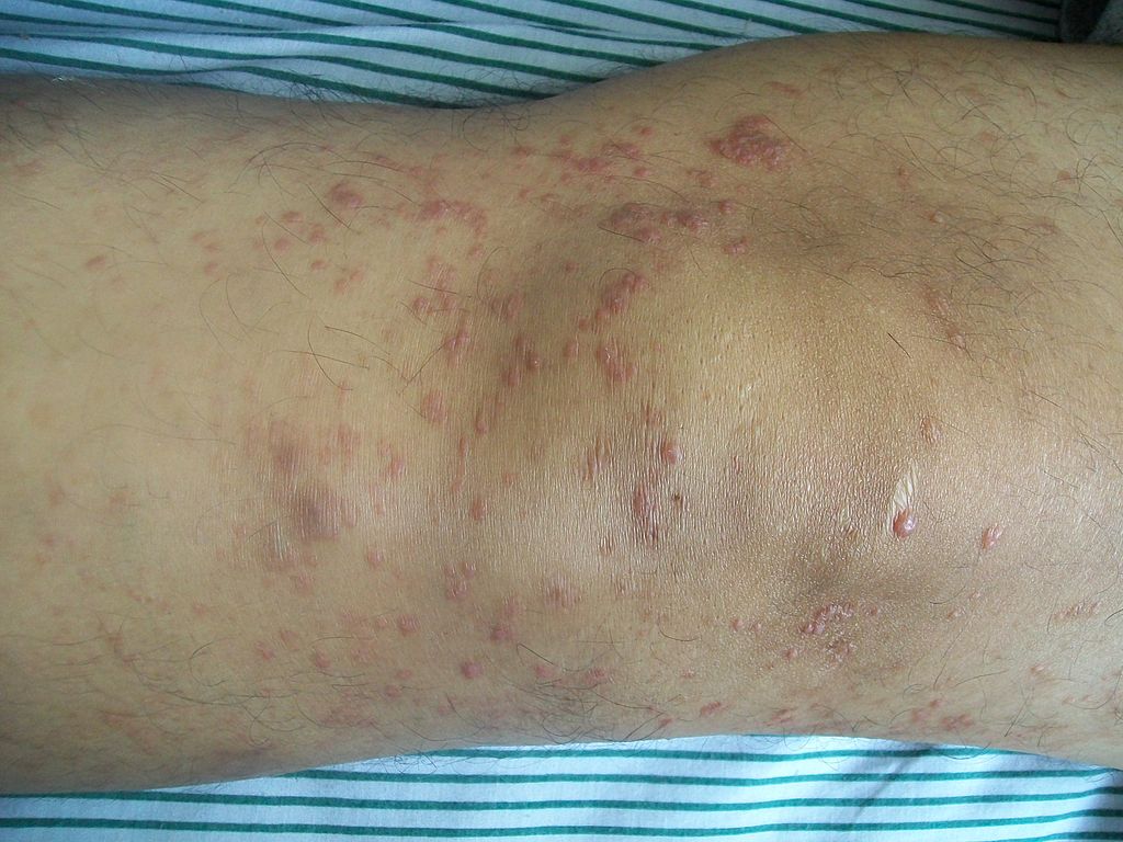
Mycosis fungoides knee


Microscopic Pathology
- Mycosis fungoides has been divided into three stages:
- Premycotic stage
- Mycotic stage
- Tumorous stage
- The premycotic stage
- Non-diagnostic and represented by chronic nonspecific dermatisis associated with psoriasiform changes in epidermis
- The mycotic stage
- Shows a polymorphous inflammatory infiltrate in the dermis that contains small numbers of frankly atypical lymphoid cells
- These cells may line up individually along the epidermal basal layer
- The latter finding if unaccompanied by spongiosis is highly suggestive of mycosis fungoides
- Tumorous stage
- Dense infiltrate of medium sized lymphocytes with cerebroid nuclei, expands the dermis
-
Sézary's disease
-
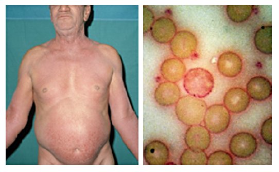
61-year-old man presented in 1972 with unrelenting pruritus of six months’ duration. On the right is his peripheral blood film stained with Periodic Acid-Schiff (PAS) showing a neoplastic T cell (Sézary cell).
-
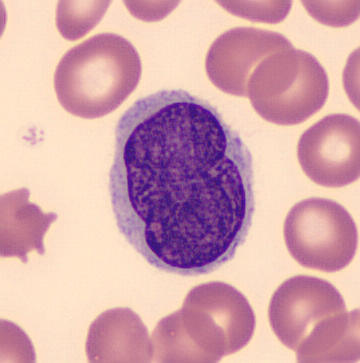
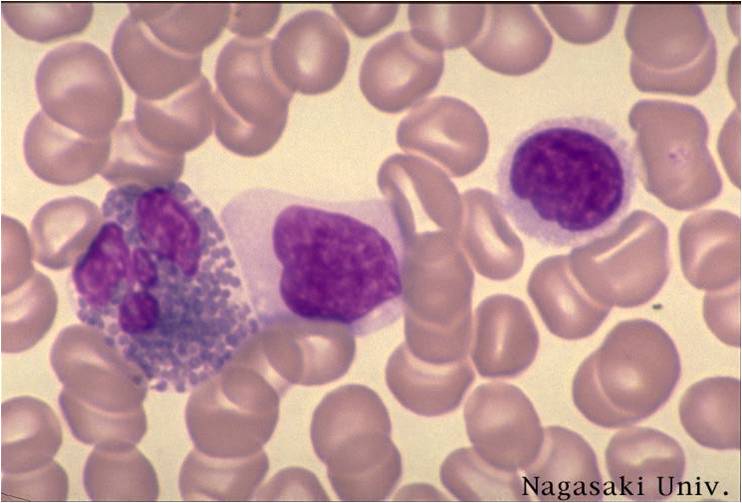
Pleomorphic abnormal T cell with the characteristic cerebriform nuclei (Peripheral blood - MGG stain)
-
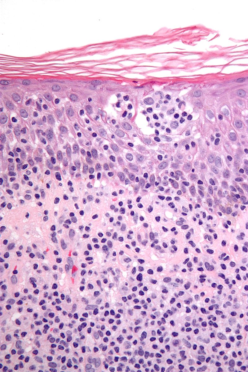
Features: Nests of lymphocytes in the epidermis; "Pautrier microabscesses". Single lymphocytes in epidermis; "lymphocyte exocytosis". Short linear arrays of lymphocytes along the basal layer of the epidermis; "epidermotropism".


