Paget's disease of the breast pathophysiology: Difference between revisions
Jump to navigation
Jump to search
| Line 24: | Line 24: | ||
*Paget cells have the same immunohistochemical staining pattern as the underlying breast cancer cells. | *Paget cells have the same immunohistochemical staining pattern as the underlying breast cancer cells. | ||
*Furthermore, they also express carcinoembryonic antigen, epithelial membrane antigen, and some mucins. | *Furthermore, they also express carcinoembryonic antigen, epithelial membrane antigen, and some mucins. | ||
*Since breast cancers associated to Paget's disease are poorly differentiated, estrogen and progesterone antigens are frequently negative | *Since breast cancers associated to Paget's disease are poorly differentiated, estrogen and progesterone antigens are frequently negative. | ||
*Mori et al found overexpression of oncogenic ras and p21 in mammary and extramammary diseases. | |||
==References== | ==References== | ||
Revision as of 20:23, 8 January 2016
|
Paget's disease of the breast Microchapters |
|
Differentiating Paget's disease of the breast from other Diseases |
|---|
|
Diagnosis |
|
Treatment |
|
Case Studies |
|
Paget's disease of the breast pathophysiology On the Web |
|
American Roentgen Ray Society Images of Paget's disease of the breast pathophysiology |
|
Directions to Hospitals Treating Paget's disease of the breast |
|
Risk calculators and risk factors for Paget's disease of the breast pathophysiology |
Editor-In-Chief: C. Michael Gibson, M.S., M.D. [1]
Overview
Pathophysiology
Gross Pathology
-
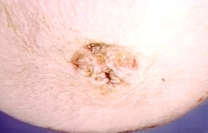
Paget's disease, nipple; Crusted eroded surface, typical lesion. Always with carcinoma.

Microscopic pathology
- Paget's disease of the breast is histopathologically characterized by epidermal Paget cells, which are malignant glandular epithelial cells with abundant and clear cytoplasm, usually containing mucin, and pleomorphic and hyperchromatic nucleus.[1]
- These cells appear organized in groups, with nest-like patterns or gland-like structures, and are preferably located in the epidermal basal layer.
- The number of cells varies from a few to large quantities; even completely replacing the epidermal cells.
- Invasion of adnexal structures can occur. Ortho-and parakeratosis may be present. The dermis displays reactive characteristics, with telangiectasia, chronic inflammation and ulceration in more advanced cases.
Immunohistochemistry
- Immunohistochemistry is very useful in Paget's disease of the breast for differential diagnoses and histogenesis.[1]
- Overexpression of the low molecular weight cytokeratins, notably CK7, and lack of expression of high molecular weight cytokeratins, such as CK10, CK14 and CK20 are observed.
- Paget cells have the same immunohistochemical staining pattern as the underlying breast cancer cells.
- Furthermore, they also express carcinoembryonic antigen, epithelial membrane antigen, and some mucins.
- Since breast cancers associated to Paget's disease are poorly differentiated, estrogen and progesterone antigens are frequently negative.
- Mori et al found overexpression of oncogenic ras and p21 in mammary and extramammary diseases.
References
- ↑ 1.0 1.1 Lopes Filho, Lauro Lourival; Lopes, Ione Maria Ribeiro Soares; Lopes, Lauro Rodolpho Soares; Enokihara, Milvia M. S. S.; Michalany, Alexandre Osores; Matsunaga, Nobuo (2015). "Mammary and extramammary Paget's disease". Anais Brasileiros de Dermatologia. 90 (2): 225–231. doi:10.1590/abd1806-4841.20153189. ISSN 1806-4841.