Multiple endocrine neoplasia type 2 CT: Difference between revisions
Jump to navigation
Jump to search
No edit summary |
(→CT) |
||
| Line 7: | Line 7: | ||
<gallery> | <gallery> | ||
Image:Pheochromocytoma CT.jpg|PheochromocytomaCase courtesy of Dr Paresh K Desai , <ref>"http://radiopaedia.org/">Radiopaedia.org</a>. From the case <a href="http://radiopaedia.org/cases/6819">rID: 6819</ref> | Image:Pheochromocytoma CT.jpg|PheochromocytomaCase courtesy of Dr Paresh K Desai , <ref>"http://radiopaedia.org/">Radiopaedia.org</a>. From the case <a href="http://radiopaedia.org/cases/6819">rID: 6819</ref> | ||
Image:Pheochromocytoma CT 2.jpg|Pheochromocytoma Case courtesy of Dr Frank Gaillard, < | Image:Pheochromocytoma CT 2.jpg|Pheochromocytoma Case courtesy of Dr Frank Gaillard, <ref>"http://radiopaedia.org/">Radiopaedia.org</a>. From the case <ref>"http://radiopaedia.org/cases/6478">rID: 6478</ref> | ||
</gallery> | </gallery> | ||
==Reference== | ==Reference== | ||
{{Reflist|2}} | {{Reflist|2}} | ||
Revision as of 19:25, 22 September 2015
|
Multiple endocrine neoplasia type 2 Microchapters |
|
Differentiating Multiple endocrine neoplasia type 2 from other Diseases |
|---|
|
Diagnosis |
|
Treatment |
|
Multiple endocrine neoplasia type 2 CT On the Web |
|
American Roentgen Ray Society Images of Multiple endocrine neoplasia type 2 CT |
|
Directions to Hospitals Treating Multiple endocrine neoplasia type 2 |
|
Risk calculators and risk factors for Multiple endocrine neoplasia type 2 CT |
Editor-In-Chief: C. Michael Gibson, M.S., M.D. [1]; Associate Editor(s)-in-Chief: Ammu Susheela, M.D. [2]
Overview
CT
- Three-dimensional single-photon emission CT (SPECT) is used for preoperative preadenoma localization.
-
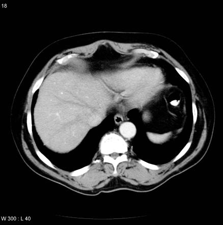
![PheochromocytomaCase courtesy of Dr Paresh K Desai , [1]](/images/7/7e/Pheochromocytoma_CT.jpg)
PheochromocytomaCase courtesy of Dr Paresh K Desai , [1]
-
Pheochromocytoma Case courtesy of Dr Frank Gaillard,
![PheochromocytomaCase courtesy of Dr Paresh K Desai , [1]](/index.php/File:Pheochromocytoma_CT.jpg)

Reference
- ↑ "http://radiopaedia.org/">Radiopaedia.org</a>. From the case <a href="http://radiopaedia.org/cases/6819">rID: 6819