Pheochromocytoma pathophysiology: Difference between revisions
No edit summary |
No edit summary |
||
| Line 5: | Line 5: | ||
On gross pathology pheochromocytoma appears as Multinodular, multicentric pattern of growth. On microscopic histopathological analysis nesting (Zellballen) pattern composed of well-defined clusters of tumour cells separated by fibrovascular stroma is a characteristic finding. | On gross pathology pheochromocytoma appears as Multinodular, multicentric pattern of growth. On microscopic histopathological analysis nesting (Zellballen) pattern composed of well-defined clusters of tumour cells separated by fibrovascular stroma is a characteristic finding. | ||
==Pathophysiology== | ==Pathophysiology== | ||
Mutations of the genes ''[[VHL]]'', ''RET'', ''NF1'', ''[[SDHB]]'' and ''[[SDHD]]'' are all known to cause familial pheochromocytoma. | |||
Traditionally it is known as the "10% tumor": | Traditionally it is known as the "10% tumor": | ||
* bilateral disease is present in approximately 10% of patients | * bilateral disease is present in approximately 10% of patients | ||
Revision as of 20:30, 2 September 2015
|
Pheochromocytoma Microchapters |
|
Diagnosis |
|---|
|
Treatment |
|
Case Studies |
|
Pheochromocytoma pathophysiology On the Web |
|
American Roentgen Ray Society Images of Pheochromocytoma pathophysiology |
|
Risk calculators and risk factors for Pheochromocytoma pathophysiology |
Editor-In-Chief: C. Michael Gibson, M.S., M.D. [1] Associate Editor(s)-in-Chief: Ahmad Al Maradni, M.D. [2]
Overview
On gross pathology pheochromocytoma appears as Multinodular, multicentric pattern of growth. On microscopic histopathological analysis nesting (Zellballen) pattern composed of well-defined clusters of tumour cells separated by fibrovascular stroma is a characteristic finding.
Pathophysiology
Mutations of the genes VHL, RET, NF1, SDHB and SDHD are all known to cause familial pheochromocytoma. Traditionally it is known as the "10% tumor":
- bilateral disease is present in approximately 10% of patients
- approximately 10% of tumours are malignant
- approximately 10% are located in chromaffin tissue outside of the adrenal gland
- Approximately 10% arise in childhood
- Approximately 10% are familial
- Approximately 10% recur after being resected
- Approximately 10% patients do not have hypertension (Campbell's Urology)These tumors can form a pattern with other endocrine gland cancers which is labeled multiple endocrine neoplasia (MEN). Pheochromocytoma may occur in patients with MEN 2 and MEN 3. VHL (Von Hippel Lindau) patients may also develop these tumors.
Tumor Location
In adults, 90% tumors are located unilaterally and are solitary, and 10% are located outside the adrenal gland. In children 50% are adrenal, while 25% are bilateral and 25% are extraadrenal. The common extradrenal locations are the abdomen, thorax and urinary bladder.
Gross Pathology
Multinodular, multicentric pattern of growth of pheochromocytoma may be seen.
-
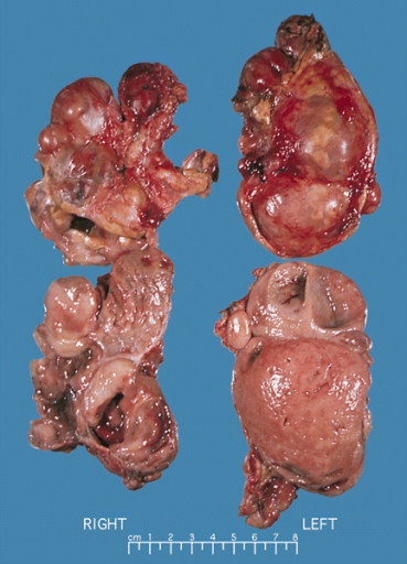
Bilateral pheochromocytoma in MEN2. Gross image.

Microscopic Pathology
Pheochromocytoma typically demonstrate a nesting (Zellballen) pattern on microscopy. This pattern is composed of well-defined clusters of tumour cells containing eosinophilic cytoplasm separated by fibrovascular stroma.
-
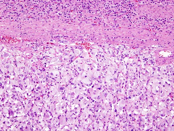
Micrograph of pheochromocytoma.
-
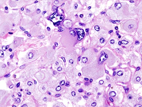
Histopathology of adrenal pheochromocytoma. Adrenectomy specimen.
-
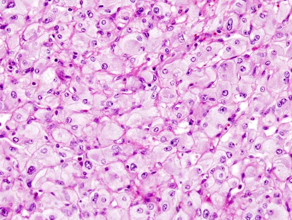
Micrograph of pheochromocytoma.
-
Micrograph of pheochromocytoma.
_histopathology.jpg)
_histopathology.jpg)
_histopathology.jpg)