Primary cutaneous amyloidosis: Difference between revisions
Kiran Singh (talk | contribs) (→Face) |
Kiran Singh (talk | contribs) (→Face) |
||
| Line 42: | Line 42: | ||
<gallery> | <gallery> | ||
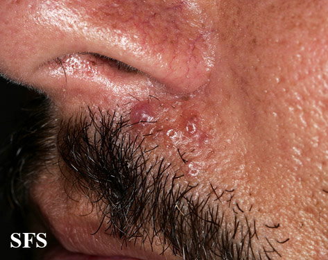
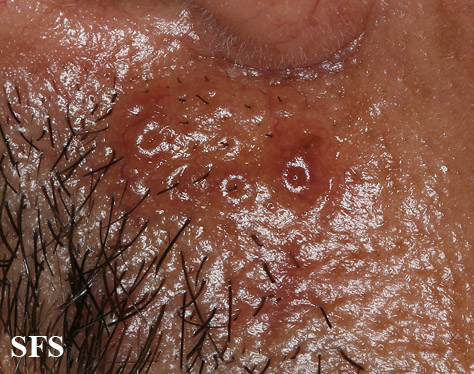
Image:Nodular amyloidosis01.jpg|Nodular amyloidosis. <SMALL><SMALL>''[http://www.atlasdermatologico.com.br/ Adapted from Dermatology Atlas.]''<ref name="Dermatology Atlas">{{Cite | Image:Nodular amyloidosis01.jpg|Nodular amyloidosis. <SMALL><SMALL>''[http://www.atlasdermatologico.com.br/ Adapted from Dermatology Atlas.]''<ref name="Dermatology Atlas">{{Cite | ||
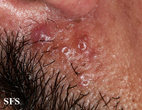
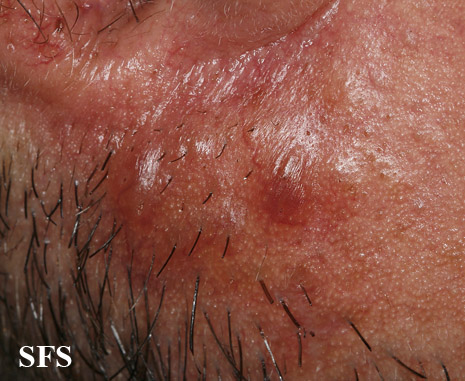
Image: | Image:Nodular amyloidosis02.jpg|Nodular amyloidosis. <SMALL><SMALL>''[http://www.atlasdermatologico.com.br/ Adapted from Dermatology Atlas.]''<ref name="Dermatology Atlas">{{Cite | ||
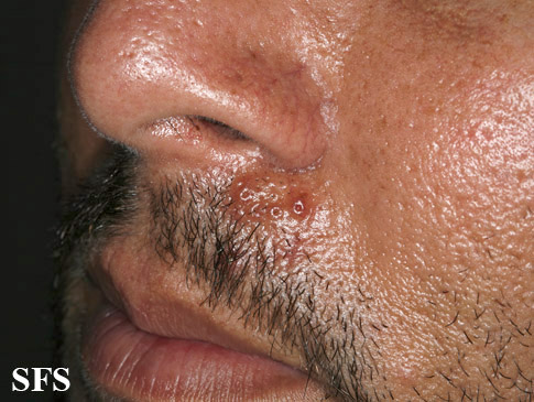
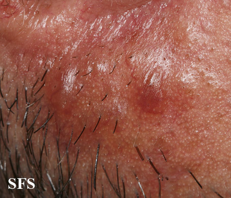
Image: | Image:Nodular amyloidosis03.jpg|Nodular amyloidosis. <SMALL><SMALL>''[http://www.atlasdermatologico.com.br/ Adapted from Dermatology Atlas.]''<ref name="Dermatology Atlas">{{Cite | ||
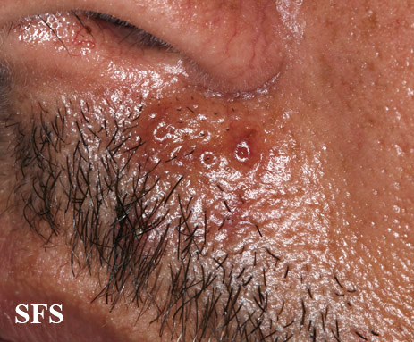
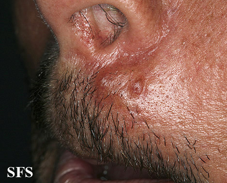
Image:Nodular amyloidosis04.jpg|Nodular amyloidosis. <SMALL><SMALL>''[http://www.atlasdermatologico.com.br/ Adapted from Dermatology Atlas.]''<ref name="Dermatology Atlas">{{Cite | Image:Nodular amyloidosis04.jpg|Nodular amyloidosis. <SMALL><SMALL>''[http://www.atlasdermatologico.com.br/ Adapted from Dermatology Atlas.]''<ref name="Dermatology Atlas">{{Cite | ||
Image:Nodular amyloidosis05.jpg|Nodular amyloidosis. <SMALL><SMALL>''[http://www.atlasdermatologico.com.br/ Adapted from Dermatology Atlas.]''<ref name="Dermatology Atlas">{{Cite | Image:Nodular amyloidosis05.jpg|Nodular amyloidosis. <SMALL><SMALL>''[http://www.atlasdermatologico.com.br/ Adapted from Dermatology Atlas.]''<ref name="Dermatology Atlas">{{Cite | ||
Image: Nodular amyloidosis06|Nodular amyloidosis. <SMALL><SMALL>''[http://www.atlasdermatologico.com.br/ Adapted from Dermatology Atlas.]''<ref name="Dermatology Atlas">{{Cite | Image:Nodular amyloidosis06|Nodular amyloidosis. <SMALL><SMALL>''[http://www.atlasdermatologico.com.br/ Adapted from Dermatology Atlas.]''<ref name="Dermatology Atlas">{{Cite | ||
Image:Nodular amyloidosis07.jpg|Nodular amyloidosis. <SMALL><SMALL>''[http://www.atlasdermatologico.com.br/ Adapted from Dermatology Atlas.]''<ref name="Dermatology Atlas">{{Cite | Image:Nodular amyloidosis07.jpg|Nodular amyloidosis. <SMALL><SMALL>''[http://www.atlasdermatologico.com.br/ Adapted from Dermatology Atlas.]''<ref name="Dermatology Atlas">{{Cite | ||
Image:Nodular amyloidosis08.jpg|Nodular amyloidosis. <SMALL><SMALL>''[http://www.atlasdermatologico.com.br/ Adapted from Dermatology Atlas.]''<ref name="Dermatology Atlas">{{Cite | Image:Nodular amyloidosis08.jpg|Nodular amyloidosis. <SMALL><SMALL>''[http://www.atlasdermatologico.com.br/ Adapted from Dermatology Atlas.]''<ref name="Dermatology Atlas">{{Cite | ||
Revision as of 02:05, 17 August 2014
| Primary cutaneous amyloidosis | |
| Classification and external resources | |
| File:Macular amyloidosis.jpg | |
|---|---|
| Macular amyloidosis, located on the right lumbar region of the back | |
| ICD-9 | 277.3 |
| OMIM | 105250 |
| DiseasesDB | 29871 |
Editor-In-Chief: C. Michael Gibson, M.S., M.D. [1];Associate Editor(s)-in-Chief: Kiran Singh, M.D. [2]
Overview
Primary cutaneous amyloidosis is a form of amyloidosis associated with oncostatin M receptor.[1][2] This type of amyloidosis has been divided into the following types:[3]:520
- Macular amyloidosis is a cutaneous condition characterized by itchy, brown, rippled macules usually located on the interscapular region of the back.[3]:521 Combined cases of lichen and macular amyloidosis are termed biphasic amyloidosis, and provide support to the theory that these two variants of amyloidosis exist on the same disease spectrum. [4]
- Lichen amyloidosis is a cutaneous condition characterized by the appearance of occasionally itchy lichenoid papules, typically appearing bilaterally on the shins.[3]:521
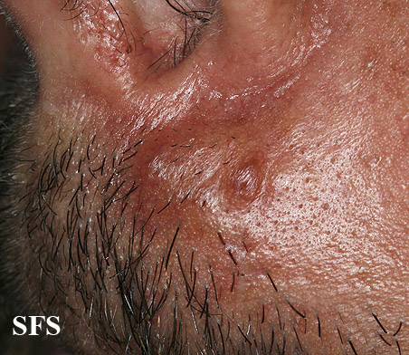
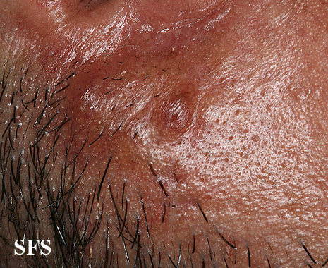
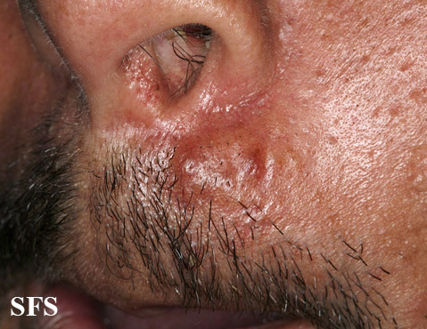
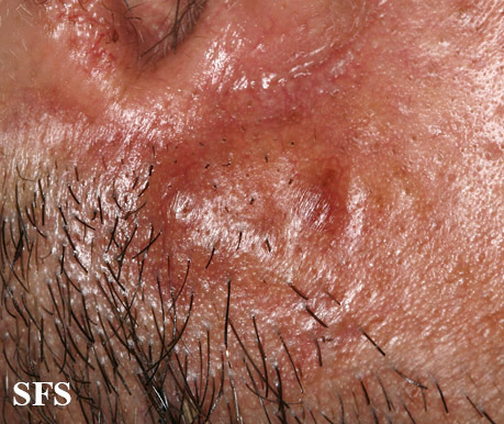
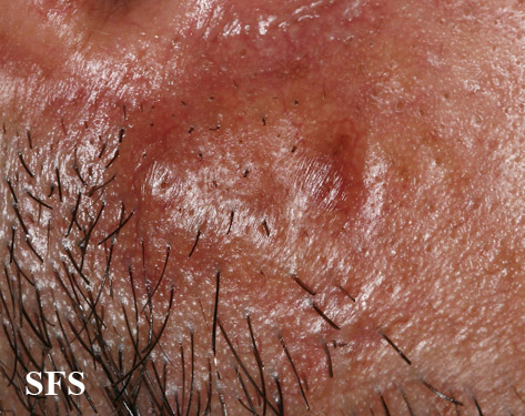
- Nodular amyloidosis is a rare cutaneous condition characterized by nodules that involve the acral areas.[3]:521
Diagnosis
Physical Examination
Skin
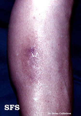
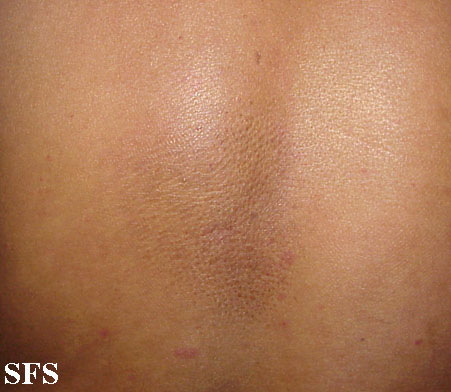
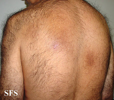
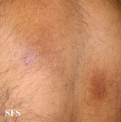
Macular Amyloidosis
Lower Extremity
-
Macular amyloidosis. Adapted from Dermatology Atlas.<ref name="Dermatology Atlas">{{Cite
Trunk
-
Macular amyloidosis. Adapted from Dermatology Atlas.<ref name="Dermatology Atlas">{{Cite
-
Macular amyloidosis. Adapted from Dermatology Atlas.<ref name="Dermatology Atlas">{{Cite
-
Macular amyloidosis. Adapted from Dermatology Atlas.<ref name="Dermatology Atlas">{{Cite
-
Macular amyloidosis. Adapted from Dermatology Atlas.<ref name="Dermatology Atlas">{{Cite
Nodular Amyloidosis
Face
-
Nodular amyloidosis. Adapted from Dermatology Atlas.<ref name="Dermatology Atlas">{{Cite
-
Nodular amyloidosis. Adapted from Dermatology Atlas.<ref name="Dermatology Atlas">{{Cite
-
Nodular amyloidosis. Adapted from Dermatology Atlas.<ref name="Dermatology Atlas">{{Cite
-
Nodular amyloidosis. Adapted from Dermatology Atlas.<ref name="Dermatology Atlas">{{Cite
-
Nodular amyloidosis. Adapted from Dermatology Atlas.<ref name="Dermatology Atlas">{{Cite
-
Nodular amyloidosis. Adapted from Dermatology Atlas.<ref name="Dermatology Atlas">{{Cite
-
Nodular amyloidosis. Adapted from Dermatology Atlas.<ref name="Dermatology Atlas">{{Cite
-
Nodular amyloidosis. Adapted from Dermatology Atlas.<ref name="Dermatology Atlas">{{Cite
-
Nodular amyloidosis. Adapted from Dermatology Atlas.<ref name="Dermatology Atlas">{{Cite
-
Nodular amyloidosis. Adapted from Dermatology Atlas.<ref name="Dermatology Atlas">{{Cite
-
Nodular amyloidosis. Adapted from Dermatology Atlas.<ref name="Dermatology Atlas">{{Cite
-
Nodular amyloidosis. Adapted from Dermatology Atlas.<ref name="Dermatology Atlas">{{Cite
-
Nodular amyloidosis. Adapted from Dermatology Atlas.<ref name="Dermatology Atlas">{{Cite
-
Nodular amyloidosis. Adapted from Dermatology Atlas.<ref name="Dermatology Atlas">{{Cite
See also
References
- ↑ "Amyloid".
- ↑ Arita K, South AP, Hans-Filho G; et al. (January 2008). "Oncostatin M receptor-beta mutations underlie familial primary localized cutaneous amyloidosis". Am. J. Hum. Genet. 82 (1): 73–80. doi:10.1016/j.ajhg.2007.09.002. PMC 2253984. PMID 18179886.
- ↑ 3.0 3.1 3.2 3.3 James, William D.; Berger, Timothy G.; et al. (2006). Andrews' Diseases of the Skin: clinical Dermatology. Saunders Elsevier. ISBN 0-7216-2921-0.
- ↑ Lichen amyloidosis of the auricular concha Craig, E. (2006) Dermatology Online Journal 12 (5): 1, University of California, Davis Department of Dermatology
|
Amyloidosis Microchapters |
|
Diagnosis |
|---|
|
Treatment |
|
Case Studies |
|
Primary cutaneous amyloidosis On the Web |
|
American Roentgen Ray Society Images of Primary cutaneous amyloidosis |
|
Risk calculators and risk factors for Primary cutaneous amyloidosis |
References
|
Amyloidosis Microchapters |
|
Diagnosis |
|---|
|
Treatment |
|
Case Studies |
|
Primary cutaneous amyloidosis On the Web |
|
American Roentgen Ray Society Images of Primary cutaneous amyloidosis |
|
Risk calculators and risk factors for Primary cutaneous amyloidosis |
