Subdural empyema: Difference between revisions
Jump to navigation
Jump to search
Gerald Chi- (talk | contribs) No edit summary |
Gerald Chi- (talk | contribs) No edit summary |
||
| Line 3: | Line 3: | ||
| Name = Subdural Empyema | | Name = Subdural Empyema | ||
| Image = Subduralempyemaandskinabscess.png | | Image = Subduralempyemaandskinabscess.png | ||
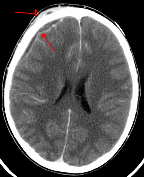
| Caption = Subdural empyema with | | Caption = Subdural empyema with skin abscess as seen on CT | ||
}} | }} | ||
'''For patient information, click [[Subdural empyema (patient information)|here]].''' | '''For patient information, click [[Subdural empyema (patient information)|here]].''' | ||
Revision as of 16:43, 27 February 2014
| Subdural Empyema | |
| Classification and external resources | |

| |
|---|---|
| Subdural empyema with skin abscess as seen on CT |
For patient information, click here.
|
Subdural empyema Microchapters |
|
Diagnosis |
|
Treatment |
|
Case Studies |
|
Subdural empyema On the Web |
|
American Roentgen Ray Society Images of Subdural empyema |
Editor-In-Chief: C. Michael Gibson, M.S., M.D. [1]
Synonyms and keywords: circumscript meningitis; pachymeningitis interna; purulent pachymeningitis; subdural abscess
Overview
Historical Perspective
Pathophysiology
Causes
Differentiating Subdural empyema from other Diseases
Epidemiology and Demographics
Risk Factors
Natural History, Complications and Prognosis
Diagnosis
History and Symptoms | Physical Examination | Laboratory Findings | Electrocardiogram | X Ray | CT | MRI | Other Diagnostic Studies
Treatment
Medical Therapy | Surgery | Prevention | Cost-Effectiveness of Therapy | Future or Investigational Therapies