Aneurysm CT: Difference between revisions
Jump to navigation
Jump to search
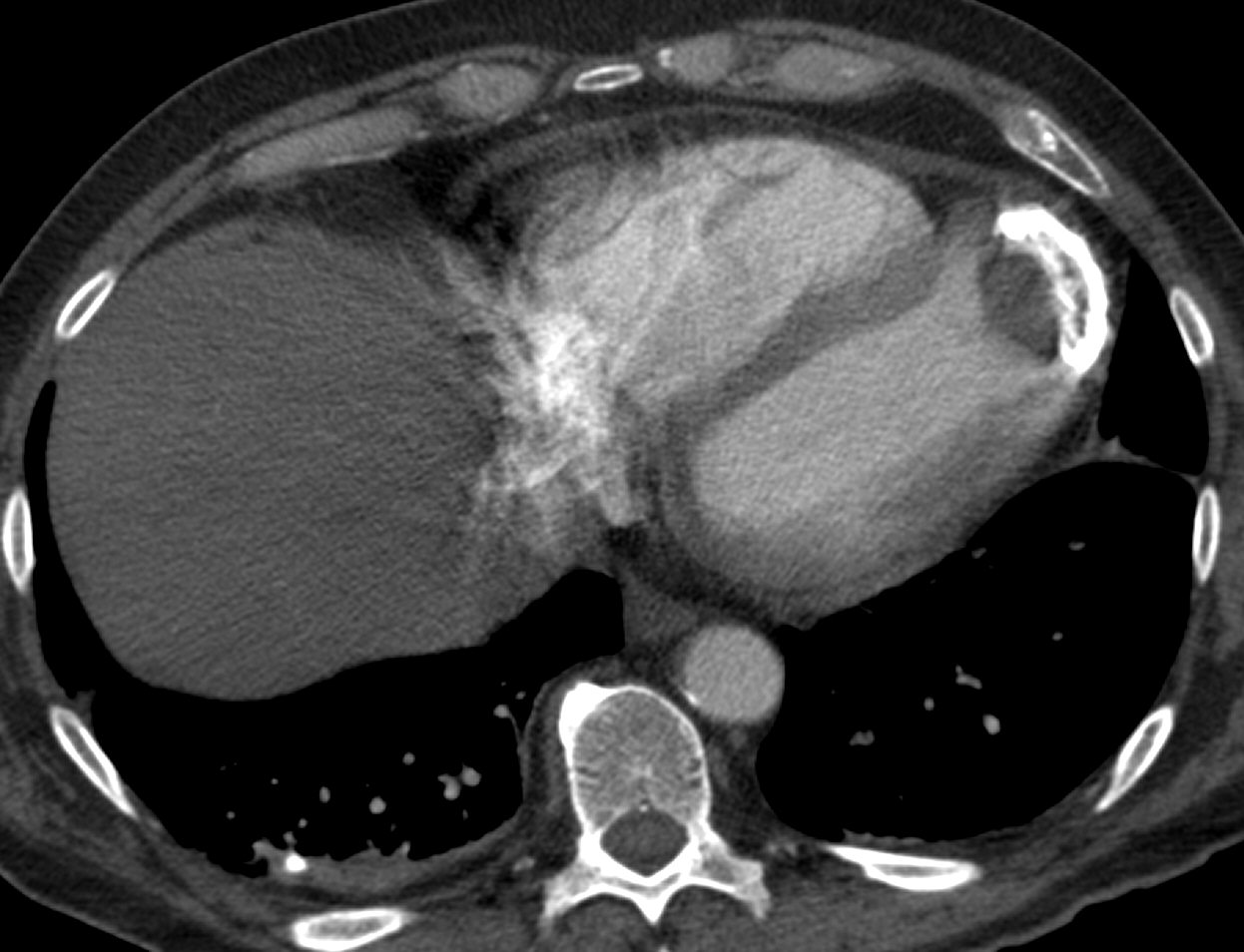
(Created page with "__NOTOC__ {{Aneurysm}} {{CMG}} ==Overview== ==CT== {| align="center" |-valign="top" | [[Image:CT.Apical aneurysm.png|thumb|Computerized Tomography image shows a left ventri...") |
No edit summary |
||
| Line 1: | Line 1: | ||
__NOTOC__ | __NOTOC__ | ||
{{Aneurysm}} | {{Aneurysm}} | ||
Please help WikiDoc by adding more content here. It's easy! Click [[Help:How_to_Edit_a_Page|here]] to learn about editing. | |||
{{CMG}} | {{CMG}} | ||
| Line 17: | Line 19: | ||
==References== | ==References== | ||
{{Reflist|2}} | {{Reflist|2}} | ||
[[Category:Needs content]] | |||
{{WH}} | {{WH}} | ||
{{WS}} | {{WS}} | ||
Revision as of 17:37, 25 August 2012
|
Aneurysm Microchapters |
Please help WikiDoc by adding more content here. It's easy! Click here to learn about editing.
Editor-In-Chief: C. Michael Gibson, M.S., M.D. [1]


