File:Acute lung abscess microscopic.png: Difference between revisions
Jump to navigation
Jump to search
Aditya Ganti (talk | contribs) (A high-power photomicrograph of the lung demonstrating a small abscess full of inflammatory cells (primarily neutrophils) (arrows). 1.There is a bacterial colony in the center of this abscess) |
(No difference)
|
Latest revision as of 17:36, 6 February 2017
A high-power photomicrograph of the lung demonstrating a small abscess full of inflammatory cells (primarily neutrophils) (arrows). 1.There is a bacterial colony in the center of this abscess
File history
Click on a date/time to view the file as it appeared at that time.
| Date/Time | Thumbnail | Dimensions | User | Comment | |
|---|---|---|---|---|---|
| current | 17:36, 6 February 2017 | 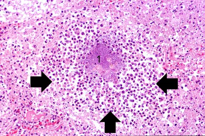 | 679 × 450 (855 KB) | Aditya Ganti (talk | contribs) | A high-power photomicrograph of the lung demonstrating a small abscess full of inflammatory cells (primarily neutrophils) (arrows). 1.There is a bacterial colony in the center of this abscess |
You cannot overwrite this file.
File usage
The following page uses this file: