Uploads by Sowminya Arikapudi
Jump to navigation
Jump to search
This special page shows all uploaded files.
| Date | Name | Thumbnail | Size | Description | Versions |
|---|---|---|---|---|---|
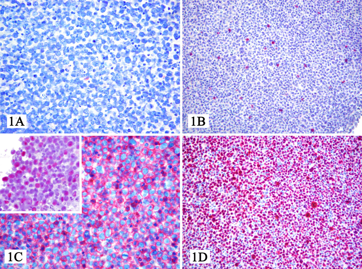
| 19:11, 23 March 2016 | Microscopic pathology of primary effusion lymphoma.jpg (file) |  |
688 KB | 1 | |
| 18:32, 10 March 2016 | Intravascular large B-cell lymphoma pathology image 9.jpg (file) |  |
41 KB | 1 | |
| 18:29, 10 March 2016 | Intravascular large B-cell lymphoma pathology image 8.jpg (file) |  |
45 KB | 1 | |
| 18:27, 10 March 2016 | Intravascular large B-cell lymphoma pathology image 7 .jpg (file) |  |
42 KB | 1 | |
| 18:25, 10 March 2016 | Intravascular large B-cell lymphoma pathology image 6.jpg (file) |  |
30 KB | 1 | |
| 18:19, 10 March 2016 | Intravascular large B-cell lymphoma pathophysiology image 5.jpg (file) |  |
37 KB | 1 | |
| 16:16, 10 March 2016 | Intravascular large B-cell lymphoma pathophysiology image 4.jpg (file) |  |
245 KB | 1 | |
| 16:04, 10 March 2016 | Intravascular large b-cell lymphoma image 3.jpg (file) |  |
244 KB | 1 | |
| 15:58, 10 March 2016 | Intravascular large b-cell lymphoma pathophysiology image 2.jpg (file) |  |
233 KB | 1 | |
| 15:53, 10 March 2016 | Intravascular large b-cell lymphoma patology image 1 .jpg (file) |  |
219 KB | 1 | |
| 15:00, 10 March 2016 | Intravascular large B-cell lymphoma CT .jpg (file) |  |
102 KB | 1 | |
| 19:28, 8 March 2016 | Subcutaneous panniculitis-like T-cell lymphoma biopsy 7.jpg (file) |  |
31 KB | 1 | |
| 19:26, 8 March 2016 | Subcutaneous panniculitis-like T-cell lymphoma biopsy 6.jpg (file) |  |
35 KB | 1 | |
| 19:24, 8 March 2016 | Subcutaneous panniculitis-like T-cell lymphoma biopsy 5.jpg (file) |  |
44 KB | 1 | |
| 19:20, 8 March 2016 | Subcutaneous panniculitis-like T-cell lymphoma biopsy 4.jpg (file) |  |
36 KB | 1 | |
| 19:16, 8 March 2016 | Subcutaneous panniculitis-like T-cell lymphoma biospy 3.jpg (file) |  |
34 KB | 1 | |
| 19:13, 8 March 2016 | Subcutaneous panniculitis-like T-cell lymphoma biopsy 2.jpg (file) |  |
42 KB | 1 | |
| 19:07, 8 March 2016 | Subcutaneous panniculitis-like T-cell lymphoma biopsy 1 .jpg (file) |  |
52 KB | 1 | |
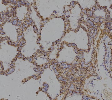
| 19:28, 7 March 2016 | Primary mediastinal large B-cell lymphoma pathology 2.jpg (file) |  |
223 KB | 1 | |
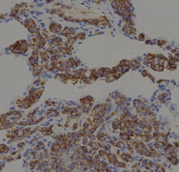
| 19:26, 7 March 2016 | Primary medistinal large b-cell lymphoma pathology 1 .jpg (file) |  |
245 KB | 1 | |
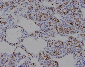
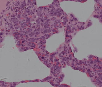
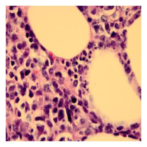
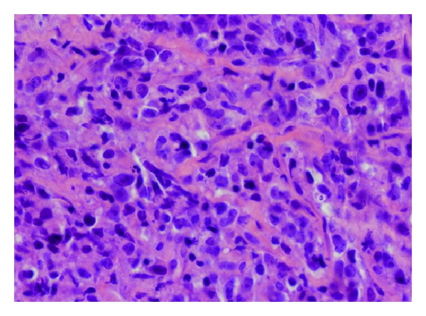
| 19:23, 7 March 2016 | Primary mediastinal large B-cell lymphoma pathology .jpg (file) |  |
202 KB | Hematoxylin and eosin (50X). Primary mediastinal B cells (PMBC) associated with delicate interstitial fibrosis. | 1 |
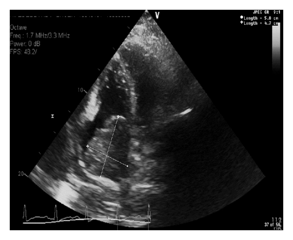
| 18:57, 7 March 2016 | Primary mediastinal large B-cell lymphoma echo.jpg (file) |  |
97 KB | A comprehensive 2 dimensional M-mode color flow and Doppler echocardiography reveals a normal left ventricular systolic function (EF 60–69%). A large right atrial mass measuring cm almost fills the right atrium and extends into the tricuspid valve... | 1 |
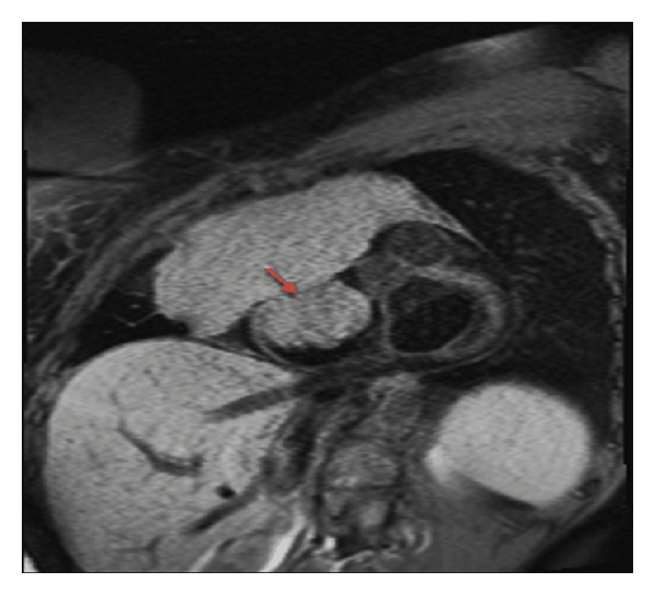
| 18:53, 7 March 2016 | Primary medistinal large B-cell lymphoma MRI .jpg (file) |  |
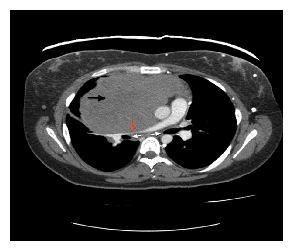
116 KB | Cardiac MRI short axis T1 at the level of mitral valve reveals a large mediastinal mass infiltrating and obliterating the SVC causing SVC obstruction. The tumor extends into the right atrium (red arrow) and invades the tricuspid valve. | 1 |
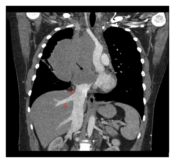
| 18:50, 7 March 2016 | Primary mediatinsl large B-cell lymphoma coronal CT scan.jpg (file) |  |
130 KB | Coronal CT scan image elucidates a mediastinal mass with extension into the right atrium (black arrow) with complete encasement and compression of the SVC. The tumor extends to the confluence of the IVC in the right atrium causing dilatation of the int... | 1 |
| 18:40, 7 March 2016 | Primary mediastinal large B-cell lymphoma CT scan 1 .jpg (file) |  |
92 KB | CT scan of the chest with contrast reveals a large lobulated anterior mediastinal solid mass (black arrow) with extension into the right hemithorax and the right atrium. There is displacement of the great vessels into the left hemithorax with significa... | 1 |
| 17:57, 2 March 2016 | Primary cutaneous follicle centre lymphoma image 10.jpg (file) |  |
179 KB | 1 | |
| 17:52, 2 March 2016 | Primary cutaneous follicle centre lymphoma image 09.jpg (file) |  |
189 KB | 1 | |
| 17:45, 2 March 2016 | Primary cutaneous follicle centre lymphoma image 08.jpg (file) |  |
194 KB | 1 | |
| 17:42, 2 March 2016 | Primary cutaneous follicle centre lymphoma image 07.jpg (file) |  |
383 KB | 1 | |
| 17:40, 2 March 2016 | Primary cutaneous follicle centre lymphoma image 06.jpg (file) |  |
873 KB | 1 | |
| 17:29, 2 March 2016 | Primary cutaneous follicle centre lymphoma image 05.jpg (file) |  |
175 KB | 1 | |
| 17:20, 2 March 2016 | Primary cutaneous follicle centre lympoma image 4.jpg (file) |  |
155 KB | 1 | |
| 14:57, 2 March 2016 | Primary cutaneous follicle centre lymphoma image 3.jpg (file) |  |
26 KB | 1 | |
| 18:38, 1 March 2016 | Primary cutaneous follicl centre lymphoma PET CT .jpg (file) |  |
392 KB | 1 | |
| 18:31, 1 March 2016 | Primary cutaneous follicle centre lymphoma image 2.jpg (file) |  |
674 KB | 1 | |
| 16:54, 1 March 2016 | Primary cutaneous follicle centre lymphoma images.jpg (file) |  |
188 KB | 1 | |
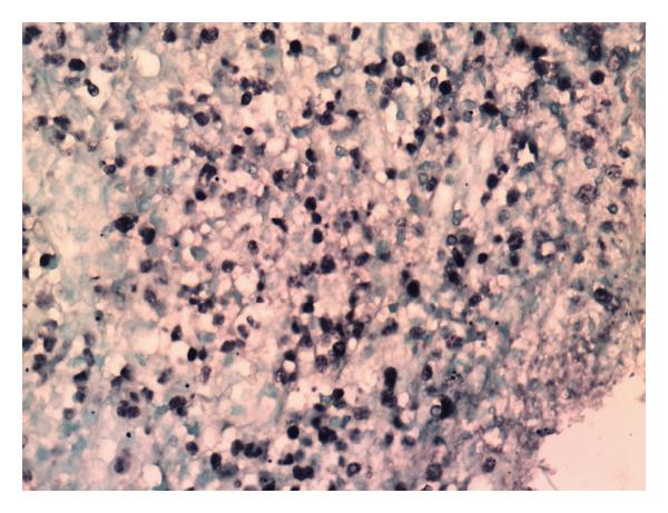
| 14:57, 24 February 2016 | Hepatosplenic T cell lymphoma bone marrow biopsy.jpg (file) |  |
188 KB | 1 | |
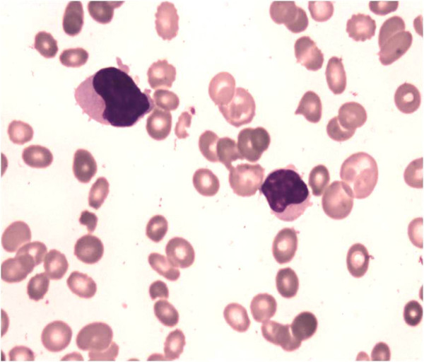
| 14:04, 24 February 2016 | Hepatosplenic T cell lymphoma peripheral blood smear.jpg (file) |  |
90 KB | 1 | |
| 14:44, 19 February 2016 | Extranodal Nk-T cell lymphoma image 2.jpg (file) |  |
53 KB | 1 | |
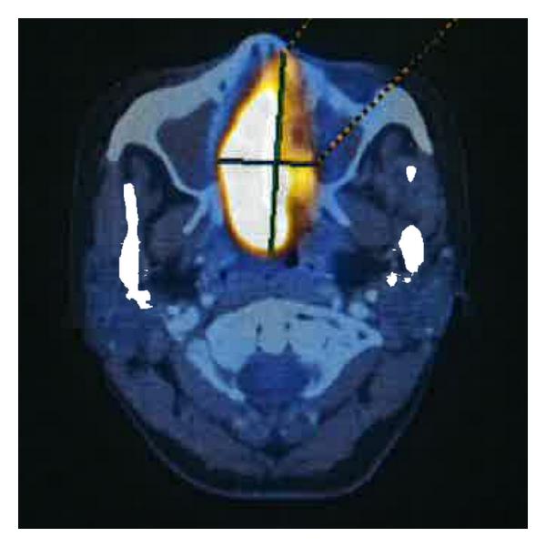
| 15:51, 18 February 2016 | Extranodal NK-T cell lymphoma PET-CT .jpg (file) |  |
28 KB | 1 | |
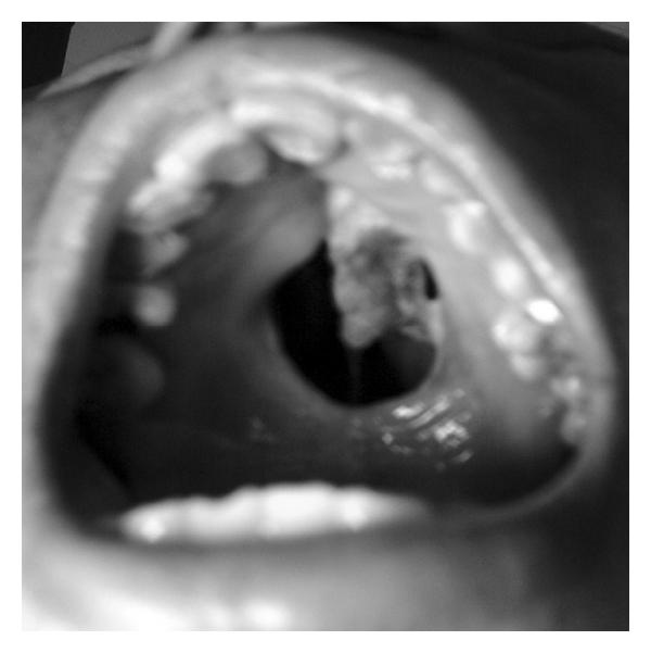
| 15:40, 18 February 2016 | Extranodal NK-T cell lymphoma image 1.jpg (file) |  |
23 KB | 1 | |
| 15:27, 18 February 2016 | Extranodal NK-T cell lymphoma.jpg (file) |  |
59 KB | 1 | |
| 16:25, 28 January 2016 | Enteropathy-associated T-cell lymphoma Immunohistochemical staining.jpg (file) |  |
309 KB | 1 | |
| 15:40, 28 January 2016 | Enteropathy-associated T-cell lymphoma Image A.jpg (file) |  |
309 KB | 1 | |
| 17:03, 27 January 2016 | Enteropathy-associated T cell lymphoma - low mag.jpg (file) |  |
331 KB | 1 | |
| 16:37, 21 January 2016 | Mycosis fungoides knee.JPG (file) |  |
135 KB | 1 | |
| 16:34, 21 January 2016 | Plaque of mycosis fungoides 1.jpg (file) |  |
200 KB | 1 | |
| 16:30, 21 January 2016 | Hem1SezaryCell2.jpg (file) |  |
20 KB | 1 | |
| 16:26, 21 January 2016 | Sézary's disease- PAS stain.jpg (file) |  |
33 KB | 1 | |
| 14:30, 21 January 2016 | Cutaneous T-cell lymphoma - very high mag.jpg (file) |  |
209 KB | 1 |