Golgi apparatus
|
WikiDoc Resources for Golgi apparatus |
|
Articles |
|---|
|
Most recent articles on Golgi apparatus Most cited articles on Golgi apparatus |
|
Media |
|
Powerpoint slides on Golgi apparatus |
|
Evidence Based Medicine |
|
Clinical Trials |
|
Ongoing Trials on Golgi apparatus at Clinical Trials.gov Trial results on Golgi apparatus Clinical Trials on Golgi apparatus at Google
|
|
Guidelines / Policies / Govt |
|
US National Guidelines Clearinghouse on Golgi apparatus NICE Guidance on Golgi apparatus
|
|
Books |
|
News |
|
Commentary |
|
Definitions |
|
Patient Resources / Community |
|
Patient resources on Golgi apparatus Discussion groups on Golgi apparatus Patient Handouts on Golgi apparatus Directions to Hospitals Treating Golgi apparatus Risk calculators and risk factors for Golgi apparatus
|
|
Healthcare Provider Resources |
|
Causes & Risk Factors for Golgi apparatus |
|
Continuing Medical Education (CME) |
|
International |
|
|
|
Business |
|
Experimental / Informatics |
Editor-In-Chief: C. Michael Gibson, M.S., M.D. [1]
Overview


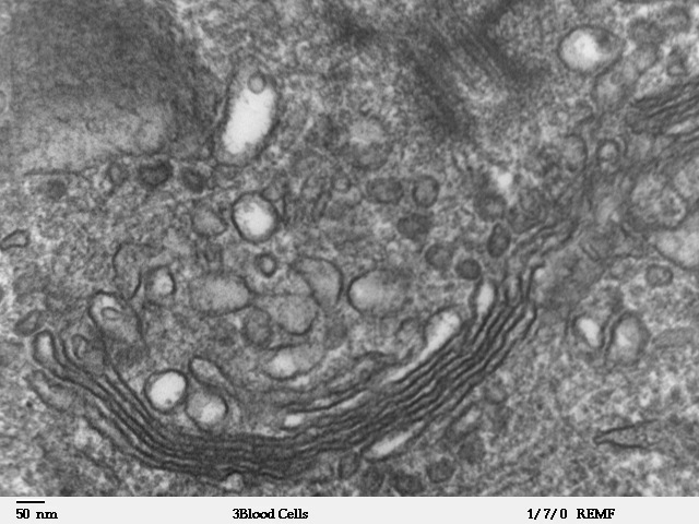
The Golgi apparatus (also called the Golgi body, Golgi complex, or dictyosome) is an organelle found in most eukaryotic cells. It was identified in 1898 by the Italian physician Camillo Golgi and was named after him. The primary function of the Golgi apparatus is to process and package the macromolecules such as proteins and lipids that are synthesised by the cell. It is particularly important in the processing of proteins for secretion. The Golgi apparatus forms a part of the endomembrane system of eukaryotic cells.
Structure
The Golgi is composed of membrane-bound sacs known as cisternae. Between five and eight are usually present; however, as many as sixty have been observed.[1]
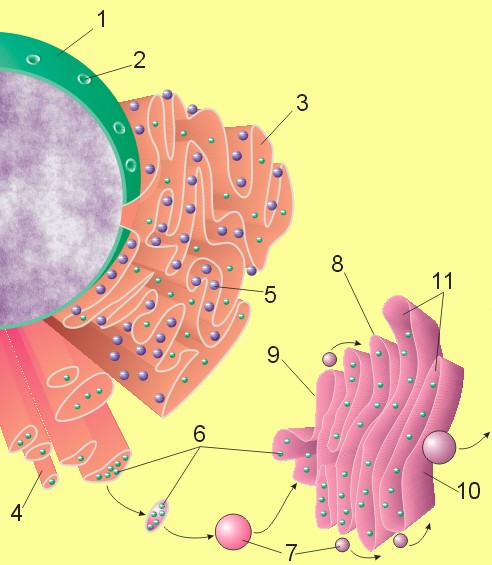
The cisternae stack has five functional regions: the cis-Golgi network, cis-Golgi, medial-Golgi, trans-Golgi, and trans-Golgi network. Vesicles from the endoplasmic reticulum (via the vesicular-tubular cluster) fuse with the cis-Golgi network and subsequently progress through the stack to the trans-Golgi network, where they are packaged and sent to the required destination. Each region contains different enzymes which selectively modify the contents depending on where they are destined to reside.[2].
Function
Cells synthesize a large number of different macromolecules required for life. The Golgi apparatus is integral in modifying, sorting, and packaging these substances for cell secretion (exocytosis) or for use within the cell. It primarily modifies proteins delivered from the rough endoplasmic reticulum, but is also involved in the transport of lipids around the cell, and the creation of lysosomes. In this respect it can be thought of as similar to a post office; it packages and labels items and then sends them to different parts of the cell.
Enzymes within the cisternae are able to modify substances by the addition of carbohydrates (glycosylation) and phosphate (phosphorylation) to them. In order to do so the Golgi transports substances such as nucleotide sugars into the organelle from the cytosol. Proteins are also labelled with a signal sequence of molecules which determine their final destination. For example, the Golgi apparatus adds a mannose-6-phosphate label to proteins destined for lysosomes.
The Golgi also plays an important role in the synthesis of proteoglycans, molecules present in the extracellular matrix of animals, and it is a major site of carbohydrate synthesis.[3]
This includes the productions of glycosaminoglycans or GAGs, long unbranched polysaccharides which the Golgi then attaches to a protein synthesized in the endoplasmic reticulum to form the proteoglycan.[4]Enzymes in the Golgi will polymerize several of these GAGs via a xylose link onto the core protein. Another task of the Golgi involves the sulfation of certain molecules passing through its lumen via sulphotranferases that gain their sulphur molecule from a donor called PAPs. This process occurs on the GAGs of proteoglycans as well as on the core protein. The level of sulfation is very important to the proteoglycans' signalling abilities as well as giving the proteoglycan its overall negative charge.[3]
The Golgi is also capable of phosphorylating molecules. To do so it transports ATP into the lumen.[5] The Golgi itself contains resident kinases, such as casein kinases. One molecule that is phosphorylated in the Golgi is Apolipoprotein, which forms a molecule known as VLDL that is a constitute of blood serum. It is thought that the phosphorylation of these molecules is important to help aid in their sorting of secretion into the blood serum.[6]
The Golgi also has a putative role in apoptosis, with several Bcl-2 family members localised there, as well as to the mitochondria. In addition a newly characterised anti-apoptotic protein, GAAP (Golgi anti-apoptotic protein), which almost exclusively resides in the Golgi, protects cells from apoptosis by an as-yet undefined mechanism (Gubser et al., 2007).
Vesicular transport
Vesicles which leave the rough endoplasmic reticulum are transported to the cis face of the Golgi apparatus, where they fuse with the Golgi membrane and empty their contents into the lumen. Once inside they are modified, sorted, and shipped towards their final destination. As such, the Golgi apparatus tends to be more prominent and numerous in cells synthesising and secreting many substances: plasma B cells, the antibody-secreting cells of the immune system, have prominent Golgi complexes.
Those proteins destined for areas of the cell other than either the endoplasmic reticulum or Golgi apparatus are moved towards the trans face, to a complex network of membranes and associated vesicles known as the trans-Golgi network (TGN).[2] This area of the Golgi is the point at which proteins are sorted and shipped to their intended destinations by their placement into one of at least three different types of vesicles, depending upon the molecular marker they carry:[2]
| Type | Description | Example |
| Exocytotic vesicles (continuous) | Vesicle contains proteins destined for extracellular release. After packaging the vesicles bud off and immediately move towards the plasma membrane, where they fuse and release the contents into the extracellular space in a process known as constitutive secretion. | Antibody release by activated plasma B cells |
| Secretory vesicles (regulated) | Vesicle contains proteins destined for extracellular release. After packaging the vesicles bud off and are stored in the cell until a signal is given for their release. When the appropriate signal is received they move towards the membrane and fuse to release their contents. This process is known as regulated secretion. | Neurotransmitter release from neurons |
| Lysosomal vesicles | Vesicle contains proteins destined for the lysosome, an organelle of degradation containing many acid hydrolases, or to lysosome-like storage organelles. These proteins include both digestive enzymes and membrane proteins. The vesicle first fuses with the late endosome, and the contents are then transferred to the lysosome via unknown mechanisms. | Digestive proteases destined for the lysosome |
Transport mechanism
The transport mechanism which proteins use to progress through the Golgi apparatus is not yet clear; however a number of hypotheses currently exist. Until recently, the vesicular transport mechanism was favoured but now more evidence is coming to light to support cisternal maturation. The two proposed models may actually work in conjunction with each other, rather than being mutually exclusive. This is sometimes referred to as the combined model. [3]
- Cisternal maturation model: the cisternae of the Golgi apparatus move by being built at the cis face and destroyed at the trans face. Vesicles from the endoplasmic reticulum fuse with each other to form a cisterna at the cis face, consequently this cisterna would appear to move through the Golgi stack when a new cisterna is formed at the cis face. This model is supported by the fact that structures larger than the transport vesicles, such as collagen rods, were observed microscopically to progress through the Golgi apparatus.[3] This was initially a popular hypothesis, but lost favour in the 1980s. Recently it has made a comeback, as laboratories at the University of Chicago and the University of Tokyo have been able to use new technology to directly observe Golgi compartments maturing.[7]. Additional evidence comes from the fact that COP1 vesicles move in the retrograde direction,. transporting ER proteins back to where they belong by recognizing a signal peptide.[8]
- Vesicular transport model: Vesicular transport views the Golgi as a very stable organelle, divided into compartments is the cis to trans direction. Membrane bound carriers transported material between the ER and Golgi and the different compartments of the Golgi.[9] Experimental evidence includes the abundance of small vesicles (known technically as shuttle vesicles) in proximity to the Golgi apparatus. Directionality is achieved by packaging proteins into either forward-moving or backward-moving (retrograde) transport vesicles, or alternatively this directionality may not be necessary as the constant input of proteins from the endoplasmic reticulum on the cis face of the Golgi would ensure flow. Irrespectively, it is likely that the transport vesicles are connected to a membrane via actin filaments to ensure that they fuse with the correct compartment.[3]
Links
References
- ↑ "Molecular Expressions Cell Biology: The Golgi Apparatus". Retrieved 2006-11-08.
- ↑ Jump up to: 2.0 2.1 2.2 Lodish (2004). Molecular Cell Biology (5th edn ed.). W.H. Freeman and Company. P0-7167-4366-3. Unknown parameter
|coauthors=ignored (help) - ↑ Jump up to: 3.0 3.1 3.2 3.3 3.4 Alberts, Bruce. Molecular Biology of the Cell. Garland Publishing. Unknown parameter
|coauthors=ignored (help) - ↑ Pyrdz, K. and K.T. Dalan, Synthesis and Sorting of Proteoglycans. Journal of Cell Science, 2000. 113: p. 193-205.
- ↑ Capasso, J., et al., Mechanism of phosphorylation in the lumen of the Golgi apparatus. Translocation of adenosine 5'-triphosphate into Golgi vesicles from rat liver and mammary gland. Journal of Biological Chemistry, 1989. 264(9): p. 5233-5240.
- ↑ Swift, L.L., Role of the Golgi Apparatus in the Phosphorylation of Apolipoprotein B. Journal of Biological Chemistry, 1996. 271(49): p. 31491-31495.
- ↑ Glick, B.S. and Malhotra, V. (1998). "The curious status of the Golgi apparatus". Cell. 95: 883–889.
- ↑ Pelham, H.R.B. and J.E. Rothman, The Debate about Transport in the Golgi - Two Sides of the Same Coin? Cell, 2000. 102: p. 713-719.
- ↑ Glick, B.S., Organisation of the Golgi apparatus. Current Opinion in Cell Biology, 2000. 12: p. 450-456.
Additional Resources
ar:جهاز كولجي bg:Апарат на Голджи ca:Aparell de Golgi cs:Golgiho aparát da:Golgiapparat de:Golgi-Apparat el:Σωμάτιο Golgi eo:Golĝi-aparato ko:골지체 hr:Golgijev aparat id:Badan Golgi is:Golgiflétta it:Apparato del Golgi he:גולג'י lb:Golgiapparat lt:Goldžio kompleksas lv:Goldži komplekss mk:Голџиев систем ms:Jasad Golgi nl:Golgi-apparaat no:Golgiapparat oc:Aparelh de Golgi simple:Golgi complex sk:Golgiho aparát sl:Golgijev aparat sr:Голџијев апарат sh:Golgijev aparat su:Awak Golgi fi:Golgin laite sv:Golgiapparaten uk:Комплекс Ґольджі