File:Primary mediastinal large B-cell lymphoma echo.jpg
Jump to navigation
Jump to search
No higher resolution available.
Primary_mediastinal_large_B-cell_lymphoma_echo.jpg (600 × 492 pixels, file size: 97 KB, MIME type: image/jpeg)
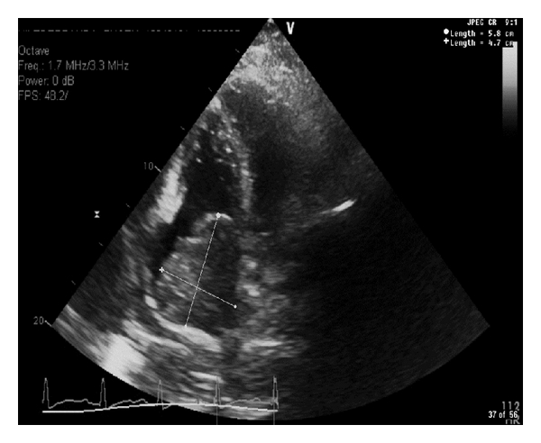
A comprehensive 2 dimensional M-mode color flow and Doppler echocardiography reveals a normal left ventricular systolic function (EF 60–69%). A large right atrial mass measuring cm almost fills the right atrium and extends into the tricuspid valve causing tricuspid regurgitation.
File history
Click on a date/time to view the file as it appeared at that time.
| Date/Time | Thumbnail | Dimensions | User | Comment | |
|---|---|---|---|---|---|
| current | 18:57, 7 March 2016 | | 600 × 492 (97 KB) | Sowminya Arikapudi (talk | contribs) | A comprehensive 2 dimensional M-mode color flow and Doppler echocardiography reveals a normal left ventricular systolic function (EF 60–69%). A large right atrial mass measuring cm almost fills the right atrium and extends into the tricuspid valve... |
You cannot overwrite this file.
File usage
There are no pages that use this file.