File:IJRI-25-109-g012.jpg
Jump to navigation
Jump to search

Size of this preview: 463 × 599 pixels. Other resolution: 539 × 697 pixels.
Original file (539 × 697 pixels, file size: 92 KB, MIME type: image/jpeg)
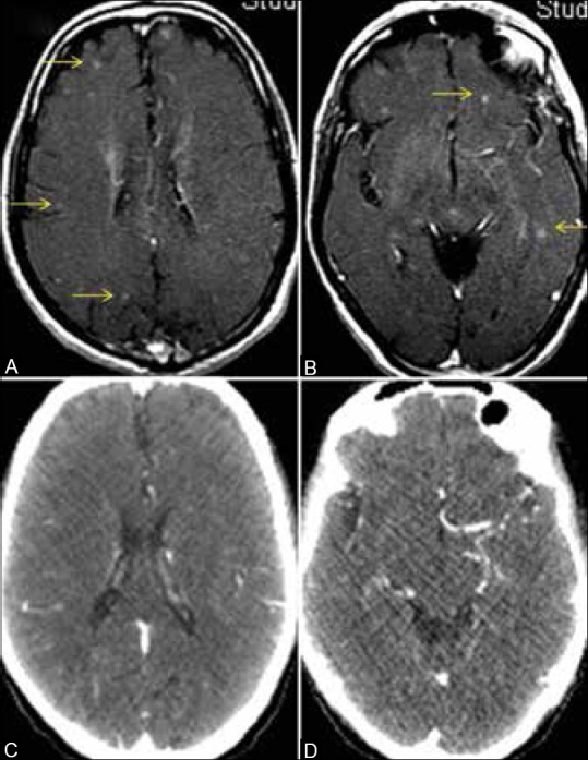
Brain metastases in asymptomatic patient, CT scan versus MRI. MRI brain in a patient of lung cancer shows multiple tiny enhancing foci scattered in the parenchyma bilaterally (arrows in A and B) suggestive of metastatic lesions. Corresponding contrast CT scan sections of the brain show no obvious lesions (C and D). Note the beam hardening effects due to bone, leading to a loss of resolution on the CT images (C and D)
File history
Click on a date/time to view the file as it appeared at that time.
| Date/Time | Thumbnail | Dimensions | User | Comment | |
|---|---|---|---|---|---|
| current | 18:19, 15 February 2018 |  | 539 × 697 (92 KB) | Dildar Hussain (talk | contribs) | Brain metastases in asymptomatic patient, CT scan versus MRI. MRI brain in a patient of lung cancer shows multiple tiny enhancing foci scattered in the parenchyma bilaterally (arrows in A and B) suggestive of metastatic lesions. Corresponding contrast... |
You cannot overwrite this file.
File usage
The following 4 pages use this file: