File:Histoplasmosis01.jpeg
Jump to navigation
Jump to search
No higher resolution available.
Histoplasmosis01.jpeg (700 × 460 pixels, file size: 48 KB, MIME type: image/jpeg)
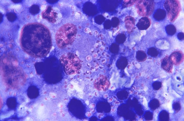
This Giemsa-stained photomicrograph reveals numerous Histoplasma capsulatum fungal organisms in their yeast-stage of development, which were seen in this liver tissue specimen, in this case of disseminated histoplasmosis.
File history
Click on a date/time to view the file as it appeared at that time.
| Date/Time | Thumbnail | Dimensions | User | Comment | |
|---|---|---|---|---|---|
| current | 04:29, 12 December 2014 | | 700 × 460 (48 KB) | Jesus Hernandez (talk | contribs) | This Giemsa-stained photomicrograph reveals numerous Histoplasma capsulatum fungal organisms in their yeast-stage of development, which were seen in this liver tissue specimen, in this case of disseminated histoplasmosis. |
You cannot overwrite this file.
File usage
The following 2 pages use this file: