File:Giardiasis06.jpeg
Jump to navigation
Jump to search
No higher resolution available.
Giardiasis06.jpeg (700 × 486 pixels, file size: 32 KB, MIME type: image/jpeg)
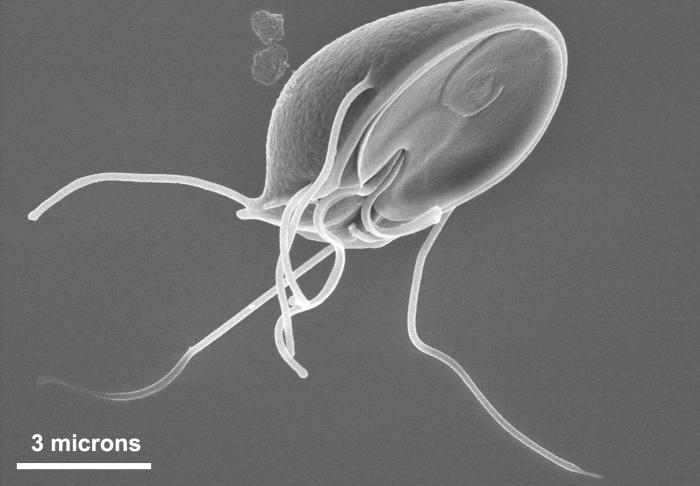
This scanning electron micrograph (SEM) clearly showed the ventral surface of a Giardia muris trophozoite. The ventral adhesive disk resembles a suction cup, where overlapping microtubules in the cytoplasm form a number-6-shaped figure. Giardia muris has four pairs of flagella that are responsible for the organism’s motility. The adhesive disk facilitates adherence of the protozoan to the intestinal surface.
File history
Click on a date/time to view the file as it appeared at that time.
| Date/Time | Thumbnail | Dimensions | User | Comment | |
|---|---|---|---|---|---|
| current | 16:20, 10 December 2014 | | 700 × 486 (32 KB) | Jesus Hernandez (talk | contribs) | This scanning electron micrograph (SEM) clearly showed the ventral surface of a Giardia muris trophozoite. The ventral adhesive disk resembles a suction cup, where overlapping microtubules in the cytoplasm form a number-6-shaped figure. Giardia muris h... |
You cannot overwrite this file.
File usage
The following page uses this file: