File:Creutzfeldt jakob02.jpeg
Jump to navigation
Jump to search
No higher resolution available.
Creutzfeldt_jakob02.jpeg (700 × 554 pixels, file size: 80 KB, MIME type: image/jpeg)
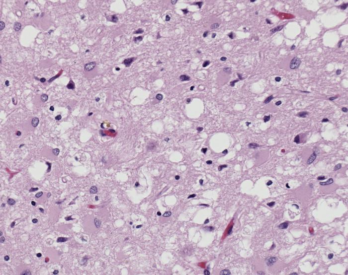
Magnified 100X, and stained with H&E (hematoxylin and eosin) staining technique, this light photomicrograph of brain tissue reveals the presence of prominent spongiotic changes in the cortex, and loss of neurons in a case of variant Creutzfeldt-Jakob disease (vCJD).
File history
Click on a date/time to view the file as it appeared at that time.
| Date/Time | Thumbnail | Dimensions | User | Comment | |
|---|---|---|---|---|---|
| current | 21:03, 4 December 2014 | | 700 × 554 (80 KB) | Jesus Hernandez (talk | contribs) | Magnified 100X, and stained with H&E (hematoxylin and eosin) staining technique, this light photomicrograph of brain tissue reveals the presence of prominent spongiotic changes in the cortex, and loss of neurons in a case of variant Creutzfeldt-Jakob d... |
You cannot overwrite this file.
File usage
The following 2 pages use this file: