File:Coronavirus02.jpeg
Jump to navigation
Jump to search
No higher resolution available.
Coronavirus02.jpeg (700 × 485 pixels, file size: 38 KB, MIME type: image/jpeg)
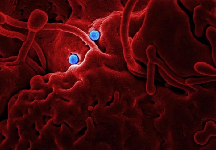
Produced by the National Institute of Allergy and Infectious Diseases (NIAID), in collaboration with Colorado State University, this highly-magnified, digitally-colorized scanning electron micrograph (SEM) reveals ultrastructural details at the site of interaction of two spherical-shaped Middle East Respiratory Syndrome Coronavirus (MERS-CoV) viral particles, colorized blue, that were on the surface of a camel epithelial cell, colorized red. Please see the Flickr link below for additional NIAID photomicrographs of the MERS-CoV.
File history
Click on a date/time to view the file as it appeared at that time.
| Date/Time | Thumbnail | Dimensions | User | Comment | |
|---|---|---|---|---|---|
| current | 20:32, 4 December 2014 | | 700 × 485 (38 KB) | Jesus Hernandez (talk | contribs) | Produced by the National Institute of Allergy and Infectious Diseases (NIAID), in collaboration with Colorado State University, this highly-magnified, digitally-colorized scanning electron micrograph (SEM) reveals ultrastructural details at the site of... |
You cannot overwrite this file.
File usage
The following page uses this file: