File:Chromoblastomycosis18.jpeg
Jump to navigation
Jump to search
No higher resolution available.
Chromoblastomycosis18.jpeg (682 × 500 pixels, file size: 57 KB, MIME type: image/jpeg)
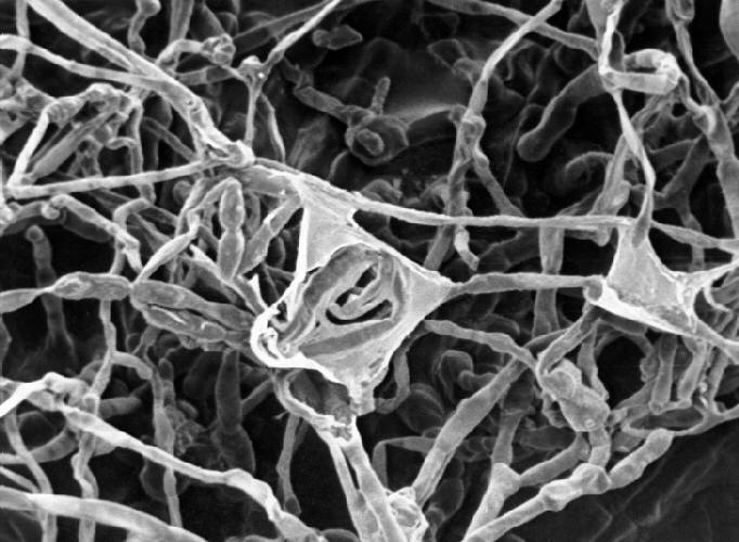
This scanning electron micrograph (SEM) reveals some of the ultrastructural morphology this pigmented, or dematiaceous mould, Xylohypha nigrescens. This mould is known to be the cause for the disease, phaeohyphomycosis, chromoblastomycosis, and mycetoma.
File history
Click on a date/time to view the file as it appeared at that time.
| Date/Time | Thumbnail | Dimensions | User | Comment | |
|---|---|---|---|---|---|
| current | 21:17, 3 December 2014 | | 682 × 500 (57 KB) | Jesus Hernandez (talk | contribs) | This scanning electron micrograph (SEM) reveals some of the ultrastructural morphology this pigmented, or dematiaceous mould, Xylohypha nigrescens. This mould is known to be the cause for the disease, phaeohyphomycosis, chromoblastomycosis, and mycetoma. |
You cannot overwrite this file.
File usage
The following page uses this file: