File:Chromoblastomycosis06.jpeg
Jump to navigation
Jump to search
No higher resolution available.
Chromoblastomycosis06.jpeg (700 × 465 pixels, file size: 40 KB, MIME type: image/jpeg)
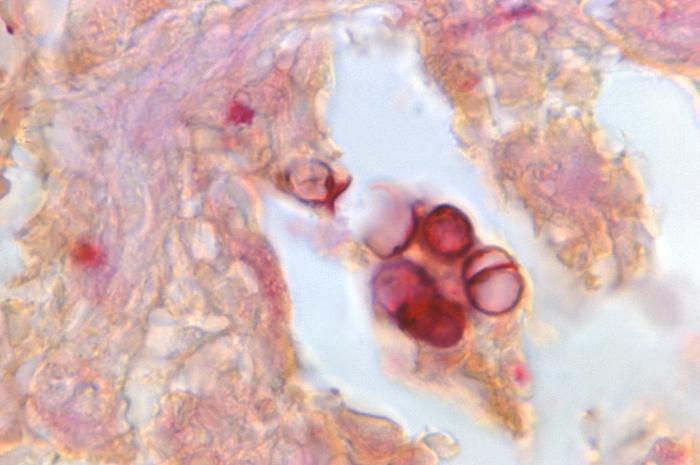
Magnified 1188X, this Gridley-stained photomicrograph revealed histopathologic changes, which were indicative of the chronic fungal disease process known as chromoblastomycosis, or chromomycosis. The tissue sample was harvested from an Indian patient. Predominantly, this infection affects the skin, or cutaneous tissues, and usually begins at the site of a puncture wound, which introduces the pathologic fungal organism into the recipient’s tissues.
File history
Click on a date/time to view the file as it appeared at that time.
| Date/Time | Thumbnail | Dimensions | User | Comment | |
|---|---|---|---|---|---|
| current | 20:53, 3 December 2014 | | 700 × 465 (40 KB) | Jesus Hernandez (talk | contribs) | Magnified 1188X, this Gridley-stained photomicrograph revealed histopathologic changes, which were indicative of the chronic fungal disease process known as chromoblastomycosis, or chromomycosis. The tissue sample was harvested from an Indian patient. ... |
You cannot overwrite this file.
File usage
The following file is a duplicate of this file (more details):
The following page uses this file: