File:Chromoblastomycosis05.jpeg
Jump to navigation
Jump to search
No higher resolution available.
Chromoblastomycosis05.jpeg (700 × 460 pixels, file size: 26 KB, MIME type: image/jpeg)
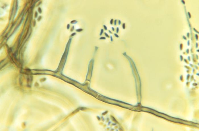
Magnified 1125X, this photomicrograph revealed morphologic details displayed by the fungal organism, Phialophora parasitica. A darkly-pigmented, filamentous fungus, which is known to be a cause of chromoblastomycosis and phaeohyphomycosis which affect the subcutaneous tissues, however, in the case of phaeohyphomycosis, many organ systems may be affected, even becoming disseminated throughout the body.
File history
Click on a date/time to view the file as it appeared at that time.
| Date/Time | Thumbnail | Dimensions | User | Comment | |
|---|---|---|---|---|---|
| current | 20:51, 3 December 2014 | | 700 × 460 (26 KB) | Jesus Hernandez (talk | contribs) | Magnified 1125X, this photomicrograph revealed morphologic details displayed by the fungal organism, Phialophora parasitica. A darkly-pigmented, filamentous fungus, which is known to be a cause of chromoblastomycosis and phaeohyphomycosis which affect ... |
You cannot overwrite this file.
File usage
The following page uses this file: