File:CT scan of brain metastasis 1.jpg
Jump to navigation
Jump to search
No higher resolution available.
CT_scan_of_brain_metastasis_1.jpg (630 × 572 pixels, file size: 31 KB, MIME type: image/jpeg)
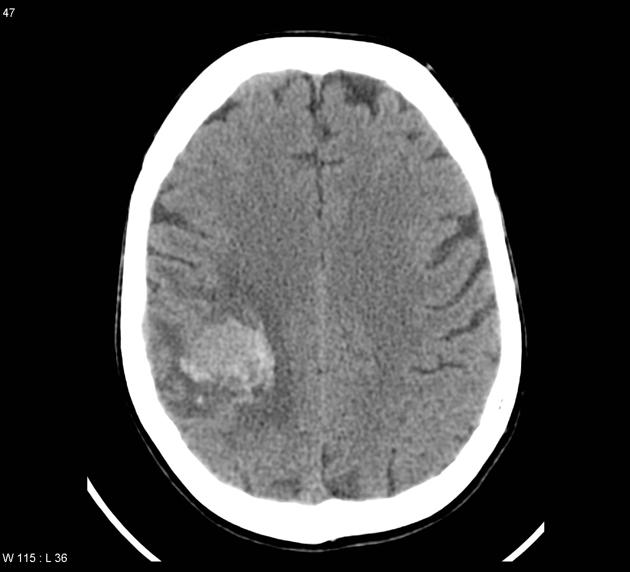
CT scan of a 60 year old patient who developed left-handed weakness which improved with corticosteroids and neurological examination revealing left pronator drift and left sided arm and leg weakness demonstrates right frontoparietal hyperdense intra-axial lesion that enhances in post contrast study. Small surrounding vasogenic edema and no significant mass effect is observed.
File history
Click on a date/time to view the file as it appeared at that time.
| Date/Time | Thumbnail | Dimensions | User | Comment | |
|---|---|---|---|---|---|
| current | 14:20, 13 November 2015 | | 630 × 572 (31 KB) | Sujit Routray (talk | contribs) | Image courtesy of Dr. Frank Gaillard. Radiopaedia (original file [http://radiopaedia.org/cases/metastatic-malignant-melanoma here]). Creative Commons BY-SA-NC |
| 14:18, 13 November 2015 |  | 630 × 572 (31 KB) | Sujit Routray (talk | contribs) | CT scan of a 60 year old patient who developed left-handed weakness which improved with corticosteroids and neurological examination revealing left pronator drift and left sided arm and leg weakness demonstrates right frontoparietal hyperdense intra-ax... | |
| 14:17, 13 November 2015 |  | 630 × 572 (31 KB) | Sujit Routray (talk | contribs) | CT scan of a 60 year old patient who developed left-handed weakness which improved with corticosteroids and neurological examination revealing left pronator drift and left sided arm and leg weakness demonstrates right frontoparietal hyperdense intra-ax... |
You cannot overwrite this file.
File usage
The following page uses this file: