File:CT image of pleural fibroma.jpg
Jump to navigation
Jump to search
No higher resolution available.
CT_image_of_pleural_fibroma.jpg (553 × 260 pixels, file size: 24 KB, MIME type: image/jpeg)
Summary
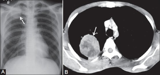
Pleural fibroma: (A) Chest radiograph showing lobulated pleural-based opacity (arrow) in right apical region; (B) axial contrast-enhanced CT scan showing heterogeneously enhancing peripheral mass lesion (arrow) in a biopsy-proven case of benign pleural fibroma,Sureka B, Thukral BB, Mittal MK, Mittal A, Sinha M. Radiological review of pleural tumors. Indian J Radiol Imaging. 2013;23(4):313–320. doi:10.4103/0971-3026.125577,https://www.ncbi.nlm.nih.gov/pmc/articles/PMC3932573/
File history
Click on a date/time to view the file as it appeared at that time.
| Date/Time | Thumbnail | Dimensions | User | Comment | |
|---|---|---|---|---|---|
| current | 15:49, 11 June 2019 | | 553 × 260 (24 KB) | Maneesha Nandimandalam (talk | contribs) | Pleural fibroma: (A) Chest radiograph showing lobulated pleural-based opacity (arrow) in right apical region; (B) axial contrast-enhanced CT scan showing heterogeneously enhancing peripheral mass lesion (arrow) in a biopsy-proven case of benign pleural... |
You cannot overwrite this file.
File usage
The following page uses this file: