File:CD2 antigen.png
Jump to navigation
Jump to search
No higher resolution available.
CD2_antigen.png (514 × 367 pixels, file size: 7 KB, MIME type: image/png)
Summary
| Description |
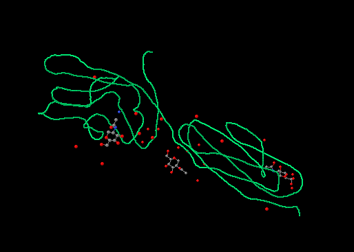
Extracytoplasmic segment of the Uman CD2 antigen, in green the protein, at the upper left the adesion domain, where is clearly recognizable the Ig structure with the three loops. There are also shown the N-ac-GLU residues where is developed a poliantennary glication based on D-man and Glu. On the right side is the proximal ig domain. |
|---|---|
| Source |
Self snapshot of a Jmol rendered pdb file 1HNF |
| Date |
12/9/06 |
| Author | |
| Permission (Reusing this file) |
GFDL CC-By-SA
|
Licensing
File history
Click on a date/time to view the file as it appeared at that time.
| Date/Time | Thumbnail | Dimensions | User | Comment | |
|---|---|---|---|---|---|
| current | 07:57, 15 July 2009 | | 514 × 367 (7 KB) | DaveBot (talk | contribs) | == Summary == {{Information |Description=Extracytoplasmic segment of the Uman CD2 antigen, in green the protein, at the upper left the adesion domain, where is clearly recognizable the Ig structure with the three loops. There are also shown the N-ac-GLU r |
You cannot overwrite this file.
File usage
There are no pages that use this file.