File:Borrelia30.jpeg
Borrelia30.jpeg (700 × 475 pixels, file size: 54 KB, MIME type: image/jpeg)
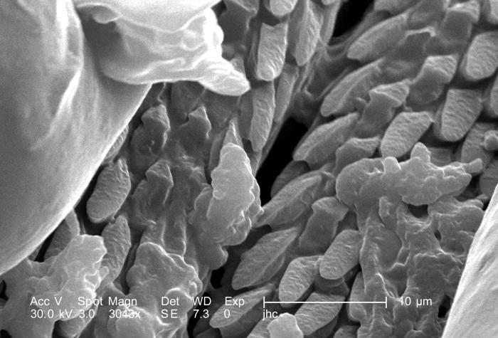
Under a magnification of 3043X, approximately 8 times greater than PHIL 9963, this scanning electron micrograph (SEM) depicted a dorsal view of an unidentified male Dermacentor sp. tick found upon a cat in the suburbs of Decatur, Georgia, which measured approximately 3.5mm from its gnathosoma (i.e., capitulum), which is where its mouthparts are located, to the distal abdominal margin (PHIL 9961). Note in PHIL 9959 and 9960, that the entire dorsum of this tick’s abdomen is covered by its tough “scutum”, or “shield”, categorizing it as a male. In female Ixodid-species ticks, the scutum only partially covers the dorsal abdomen. Seen clearly in this image is the foliate covering of the tick’s skin-piercing hypostome, which is located in what appears to be a trough between its pedipalps.
File history
Click on a date/time to view the file as it appeared at that time.
| Date/Time | Thumbnail | Dimensions | User | Comment | |
|---|---|---|---|---|---|
| current | 15:19, 26 November 2014 | | 700 × 475 (54 KB) | Jesus Hernandez (talk | contribs) | Under a magnification of 3043X, approximately 8 times greater than PHIL 9963, this scanning electron micrograph (SEM) depicted a dorsal view of an unidentified male Dermacentor sp. tick found upon a cat in the suburbs of Decatur, Georgia, which measure... |
You cannot overwrite this file.
File usage
The following page uses this file: