File:Borrelia02.jpeg
Original file (700 × 714 pixels, file size: 77 KB, MIME type: image/jpeg)
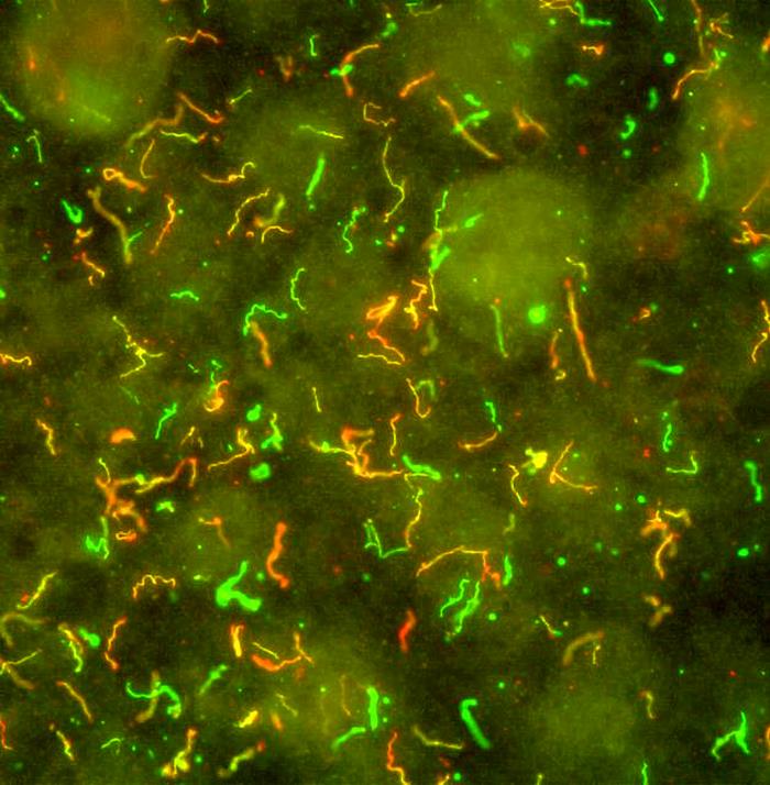
Produced by the National Institute of Allergy and Infectious Diseases (NIAID), this photomicrographic montage was created by combining two slides processed using the immunofluorescent antibody technique (IFA). One slide was used to identify spirochetes that express outer surface protein D, resulting in yellow- and red-colored organisms. Again using IFA on the second slide, spirochetes were labeled with an anti-B. burgdorferi antibody, producing organisms that had stained a glowing green color. The two slides were then combined producing this B. burgdorferi multicolored image.
File history
Click on a date/time to view the file as it appeared at that time.
| Date/Time | Thumbnail | Dimensions | User | Comment | |
|---|---|---|---|---|---|
| current | 14:36, 26 November 2014 | | 700 × 714 (77 KB) | Jesus Hernandez (talk | contribs) | Produced by the National Institute of Allergy and Infectious Diseases (NIAID), this photomicrographic montage was created by combining two slides processed using the immunofluorescent antibody technique (IFA). One slide was used to identify spirochetes... |
You cannot overwrite this file.
File usage
The following page uses this file: