File:Blastomycosis21.jpeg
Jump to navigation
Jump to search
No higher resolution available.
Blastomycosis21.jpeg (700 × 462 pixels, file size: 44 KB, MIME type: image/jpeg)
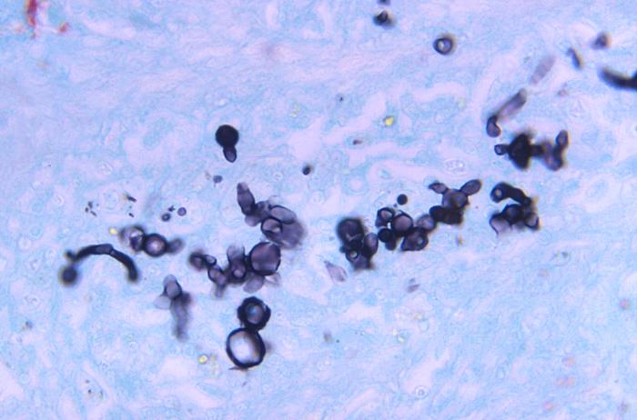
Magnified 500X, this Gamori-stained photomicrograph of a canine liver tissue specimen, revealed the presence of budding Blastomyces dermatitidis fungal cells of various sizes. Note the accompanying filaments, or mycelium.
File history
Click on a date/time to view the file as it appeared at that time.
| Date/Time | Thumbnail | Dimensions | User | Comment | |
|---|---|---|---|---|---|
| current | 21:40, 24 November 2014 | | 700 × 462 (44 KB) | Jesus Hernandez (talk | contribs) | Magnified 500X, this Gamori-stained photomicrograph of a canine liver tissue specimen, revealed the presence of budding Blastomyces dermatitidis fungal cells of various sizes. Note the accompanying filaments, or mycelium. |
You cannot overwrite this file.
File usage
The following page uses this file: