File:Blastomycosis16.jpeg
Jump to navigation
Jump to search
Size of this preview: 636 × 600 pixels. Other resolution: 700 × 660 pixels.
Original file (700 × 660 pixels, file size: 48 KB, MIME type: image/jpeg)
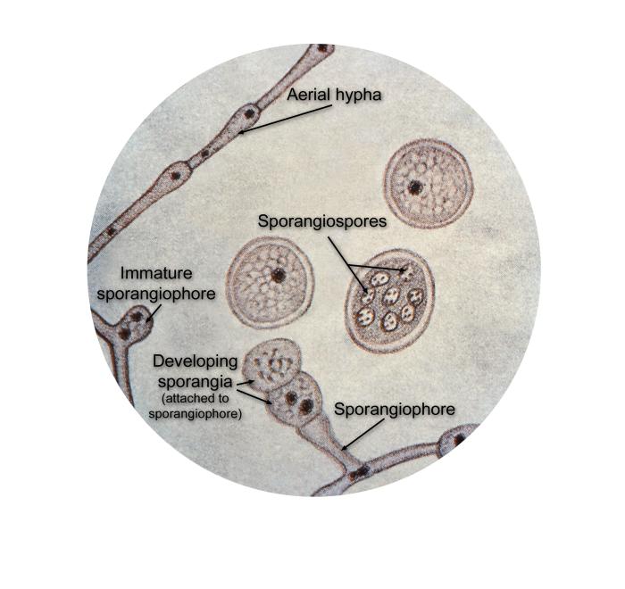
This illustration depicts the ultrastructural details found in the dimorphic fungal organism, Blastomyces dermatitidis including the organism’s aerial hypha, developing sporangia, which would eventually contain mature sporangiospores, and the sporangiophore, which gives rise to the developing sporangia.
File history
Click on a date/time to view the file as it appeared at that time.
| Date/Time | Thumbnail | Dimensions | User | Comment | |
|---|---|---|---|---|---|
| current | 21:32, 24 November 2014 | | 700 × 660 (48 KB) | Jesus Hernandez (talk | contribs) | This illustration depicts the ultrastructural details found in the dimorphic fungal organism, Blastomyces dermatitidis including the organism’s aerial hypha, developing sporangia, which would eventually contain mature sporangiospores, and the sporang... |
You cannot overwrite this file.
File usage
The following page uses this file: