File:Blastomycosis05.jpeg
Jump to navigation
Jump to search
No higher resolution available.
Blastomycosis05.jpeg (700 × 459 pixels, file size: 70 KB, MIME type: image/jpeg)
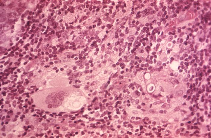
This hematoxylin-eosin (H&E)-stained photomicrograph reveals some of the ultrastructural histopathology in an dermal skin tissue specimen in a patient with an intradermal keloidean blastomycosis infection, which was caused by the fungus, Blastomyces dermatitidis. In this particular section, you’ll note presence of a parasitized multinucleated giant cell.
File history
Click on a date/time to view the file as it appeared at that time.
| Date/Time | Thumbnail | Dimensions | User | Comment | |
|---|---|---|---|---|---|
| current | 21:19, 24 November 2014 | | 700 × 459 (70 KB) | Jesus Hernandez (talk | contribs) | This hematoxylin-eosin (H&E)-stained photomicrograph reveals some of the ultrastructural histopathology in an dermal skin tissue specimen in a patient with an intradermal keloidean blastomycosis infection, which was caused by the fungus, Blastomyces de... |
You cannot overwrite this file.
File usage
The following page uses this file: