File:Blastomycosis02.jpeg
Jump to navigation
Jump to search
No higher resolution available.
Blastomycosis02.jpeg (700 × 570 pixels, file size: 72 KB, MIME type: image/jpeg)
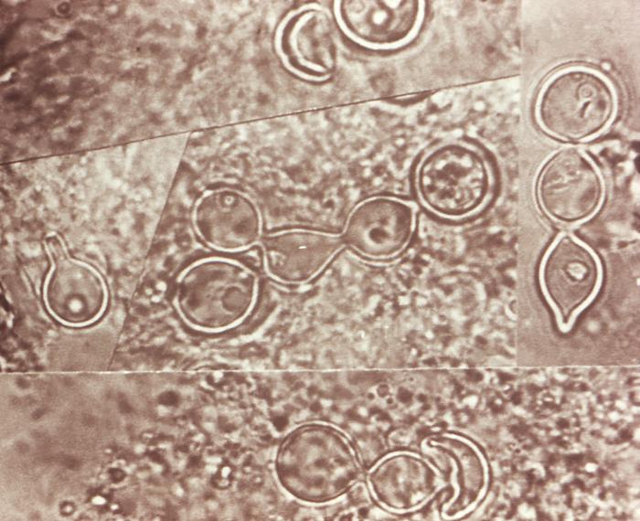
This composite photomicrograph reveals some of the ultrastructural histopathology in a tissue specimen from a patient with a keloidean blastomycosis infection, which was caused by the fungus, Blastomyces dermatitidis. The specimen originated from tissue scraping samplings. It’s important to note the abundance of large budding cells that had been configured in chains.
File history
Click on a date/time to view the file as it appeared at that time.
| Date/Time | Thumbnail | Dimensions | User | Comment | |
|---|---|---|---|---|---|
| current | 21:14, 24 November 2014 | | 700 × 570 (72 KB) | Jesus Hernandez (talk | contribs) | This composite photomicrograph reveals some of the ultrastructural histopathology in a tissue specimen from a patient with a keloidean blastomycosis infection, which was caused by the fungus, Blastomyces dermatitidis. The specimen originated from tissu... |
You cannot overwrite this file.
File usage
The following page uses this file: