File:Anaplasma phagocytophilum02.jpeg
Jump to navigation
Jump to search
No higher resolution available.
Anaplasma_phagocytophilum02.jpeg (700 × 475 pixels, file size: 68 KB, MIME type: image/jpeg)
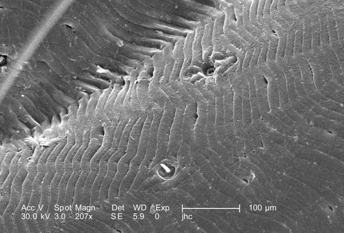
Under a magnification of 207X, this scanning electron micrographic (SEM) image depicted a dorsal view of an unidentified engorged female tick which had been extracted from the skin of a pet cat while in the process of obtaining its blood meal. Seen here, is an enlarged view of the exoskeletal surface of the engorged abdomen, which reveals the wrinkled, expandable tissue that enables the tick to ingest huge quantities of blood.
File history
Click on a date/time to view the file as it appeared at that time.
| Date/Time | Thumbnail | Dimensions | User | Comment | |
|---|---|---|---|---|---|
| current | 05:11, 12 December 2014 | | 700 × 475 (68 KB) | Jesus Hernandez (talk | contribs) | Under a magnification of 207X, this scanning electron micrographic (SEM) image depicted a dorsal view of an unidentified engorged female tick which had been extracted from the skin of a pet cat while in the process of obtaining its blood meal. Seen her... |
You cannot overwrite this file.
File usage
The following page uses this file: