File:Amebiasis06.jpeg
Amebiasis06.jpeg (700 × 525 pixels, file size: 64 KB, MIME type: image/jpeg)
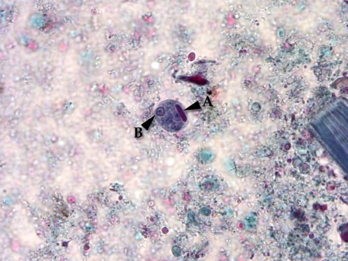
Using a trichrome stain, this photomicrograph depicted a cyst of the single-celled parasite, Entamoeba histolytica. Stained a blue color, the cyst, see here in the center of the micrograph, is one of the life cycle phases through which a protozoan organism passes as it matures. In this phase, due to the protective cyst wall, the organism is extremely resilient to the elements and is able to survive from days to weeks in the external environment. The cyst represents the highly infective phase of the life cycle. Note the presence of an elongated, blunt ended chromatoid body within the cyst “A”, and a well-defined nucleus “B”.
File history
Click on a date/time to view the file as it appeared at that time.
| Date/Time | Thumbnail | Dimensions | User | Comment | |
|---|---|---|---|---|---|
| current | 16:43, 20 November 2014 | | 700 × 525 (64 KB) | Jesus Hernandez (talk | contribs) | Using a trichrome stain, this photomicrograph depicted a cyst of the single-celled parasite, Entamoeba histolytica. Stained a blue color, the cyst, see here in the center of the micrograph, is one of the life cycle phases through which a protozoan orga... |
You cannot overwrite this file.
File usage
The following page uses this file: