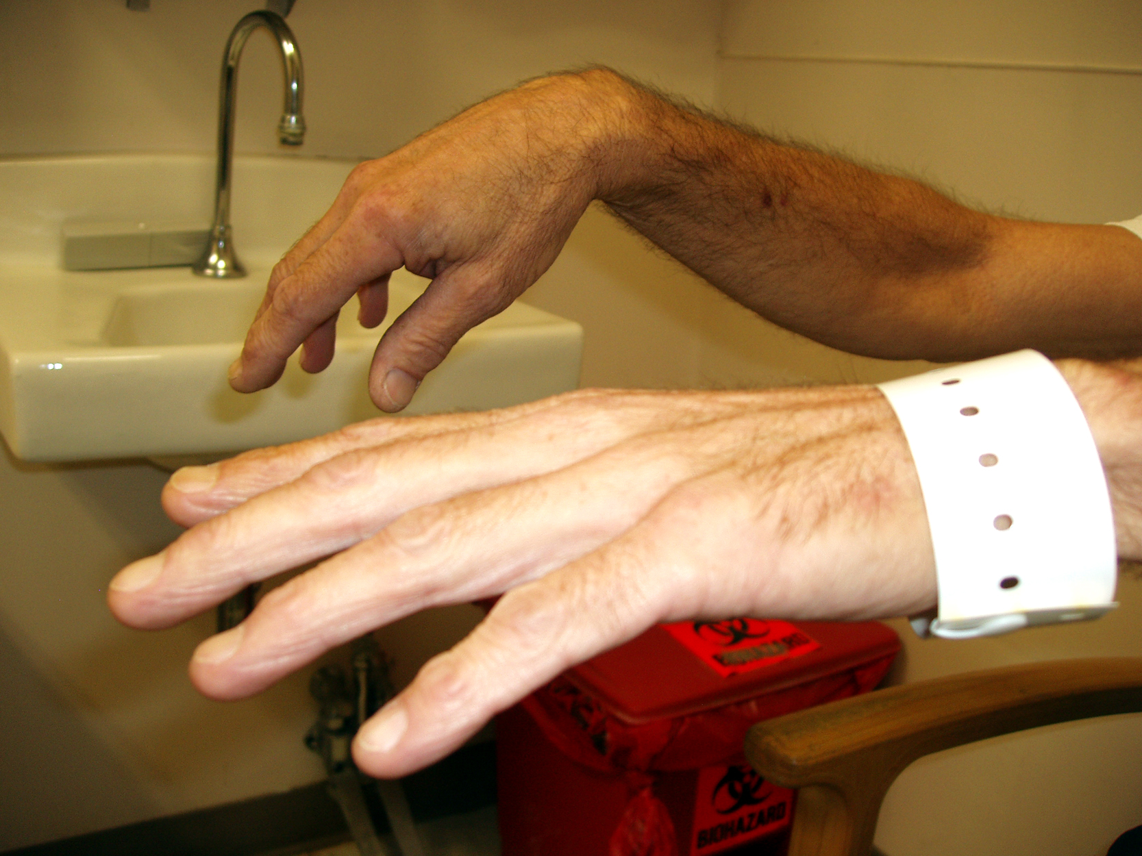
Wrist drop
For patient information, click here
| Wrist drop | |
 | |
|---|---|
| Radial Nerve Palsy: Note inability of patient to extend right wrist. (Image courtesy of Charlie Goldberg, M.D.) |
|
Wrist Drop Microchapters |
|
Diagnosis |
|---|
|
Treatment |
|
Case Studies |
|
Wrist drop On the Web |
|
American Roentgen Ray Society Images of Wrist drop |
Editor-In-Chief: C. Michael Gibson, M.S., M.D. [1]; Associate Editor-In-Chief: Cafer Zorkun, M.D., Ph.D. [2]
Synonyms and keywords: Radial nerve palsy
Overview
Wrist drop, also known as radial nerve palsy, is a condition where a person can not extend their wrist and it hangs flaccidly. To demonstrate wrist drop, hold your arm out in front of you with your forearm parallel to the floor. With the back of your hand facing the ceiling (i.e. pronated), let your hand hang limply so that your fingers point downward. A person with wrist drop would be unable to move from this position to one in which the fingers are pointing up towards the ceiling.
Anatomy of the forearm
In anatomical parlance, the forearm is the part of the body which extends from the elbow to the wrist and is not to be confused with the arm which extends from the shoulder to the elbow. The extensor muscles in the forearm are extensor carpi ulnaris, extensor digiti minimi, extensor digitorum, extensor indicis, extensor pollicis longus, extensor pollicis brevis, extensor carpi radialis brevis, extensor carpi radialis longus. These extensor muscles are supplied by the radial nerve. Other muscles in the forearm also innervated by the radial nerve are brachioradialis, supinator and abductor pollicis longus. Note that all these muscles are situated in the posterior half of the forearm (posterior when in the anatomical position).
Differential diagnosis of causes of wrist drop
Wrist extension is achieved by muscles in the forearm contracting, pulling on tendons that attach distal to (beyond) the wrist. If the tendons, the muscles, or the nerves supplying these muscles, are not working as they should be, wrist drop may occur. The following situations may result in wrist drop:
Stab wounds to the chest at or below the clavicle may result in wrist drop. The radial nerve is the terminal branch of the posterior cord of the brachial plexus. A stab wound may damage the posterior cord and result in neurological deficeits including an inability to abduct the shoulder beyond 15 degrees, an inability to extend the forearm, reduced ability to supinate the hand, reduced ability to abduct the thumb and sensory loss to the posterior surface of the arm and hand.
The radial nerve can be damaged if the humerus (the bone of the arm) is broken, because it runs through the radial groove on the lateral border of this bone.
Wrist drop is also associated with lead poisoning because of the effect of lead on the radial nerve.[1]
Persistent injury to the nerve is also a common cause through either repetitive motion or by applying pressure externally along the route of the radial nerve as in the prolonged use of crutches or extended leaning on the elbows.
Diagnosis
The workup for wrist drop frequently includes nerve conduction velocity studies to isolate and confirm the radial nerve as the source of the problem. Plain films can help identify bone spurs and fractures that may have injured the nerve. Sometimes MRI imaging is required to differentiate subtle causes.
Treatment
Initial management includes splinting of the wrist for support along with occupational or physical therapy. In some cases surgical removal of bone spurs or other anatomical defects that may be impinging on the nerve might be warranted.
See also
References
- ↑ Dedeken P, Louw V, Vandooren AK, Geert V, Goossens W, Dubois B (2006). "Plumbism or lead intoxication mimicking an abdominal tumor". Journal of general internal medicine : official journal of the Society for Research and Education in Primary Care Internal Medicine. 21 (6): C1–3. doi:10.1111/j.1525-1497.2006.00328.x. PMID 16808730.
Template:Diseases of the musculoskeletal system and connective tissue