Wide complex tachycardia differential diagnosis
|
Wide complex tachycardia Microchapters |
|
Diagnosis |
|---|
|
Treatment |
|
Case Studies |
|
Wide complex tachycardia differential diagnosis On the Web |
|
American Roentgen Ray Society Images of Wide complex tachycardia differential diagnosis |
|
Risk calculators and risk factors for Wide complex tachycardia differential diagnosis |
Editor-In-Chief: C. Michael Gibson, M.S., M.D. [1]
Overview
Differential Diagnosis
Differentiating VT from SVT
Brugada Criteria
| Absence of an RS complex in all precordial leads? | Yes? VT (SN=0.21 SP=1.0) | ||||||||||||||||||||||||||||||||||||||||
| No? | |||||||||||||||||||||||||||||||||||||||||
| R to S interval>100 ms in one precordial lead? | Yes? VT (SN=0.21 SP=1.0) | ||||||||||||||||||||||||||||||||||||||||
| No? | |||||||||||||||||||||||||||||||||||||||||
| AV dissociation? | Yes? VT (SN=0.82 SP=0.98) | ||||||||||||||||||||||||||||||||||||||||
| No? | |||||||||||||||||||||||||||||||||||||||||
| Morphology criteria for VT present both in precordial leads V1, V2 and V6? | Yes? VT (SN=0.987 SP=0.965) | ||||||||||||||||||||||||||||||||||||||||
| No? | |||||||||||||||||||||||||||||||||||||||||
| SVT (SN=0.965 SP=0.987) | |||||||||||||||||||||||||||||||||||||||||
Vereckei Criteria
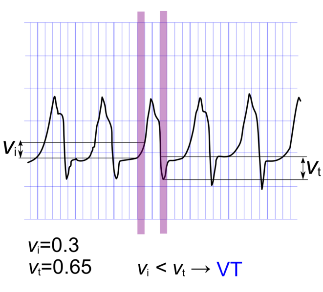
An algorithm has been proposed by Vereckei and colleagues. In addition to to do the traditional criteria, the voltage change on the EKG is used as a final discriminatory criteria. In this method, the voltage change during the initial 40 ms (v(i)) and the terminal 40 ms (v(t)) of the same QRS complex is used to estimate the (v(i)) and terminal (v(t)) ventricular activation velocity ratio (v(i)/v(t)). A v(i)/v(t) > 1 suggests SVT and a v(i)/v(t) ≤ 1 suggests VT.[1]
| AV dissociation present? | Yes? VT | ||||||||||||||||||||||||||||||||||||||||
| No? | |||||||||||||||||||||||||||||||||||||||||
| Initial R wave in aVR present? | Yes? VT | ||||||||||||||||||||||||||||||||||||||||
| No? | |||||||||||||||||||||||||||||||||||||||||
| QRS morphology unlike BBB or FB | Yes? VT | ||||||||||||||||||||||||||||||||||||||||
| No? | |||||||||||||||||||||||||||||||||||||||||
| Vi/Vt≤1? | Yes? VT | ||||||||||||||||||||||||||||||||||||||||
| No? | |||||||||||||||||||||||||||||||||||||||||
| SVT | |||||||||||||||||||||||||||||||||||||||||
Calculation of Vi/Vt
Shown below is an image demonstrating the method used to calculate Vi/Vt. In this tracing, Vi/Vt is < 1 is suggestive of ventricular tachycardia according to Vereckei criteria.
ACC Algorithm for Distinguishing SVT from VT
| Wide QRS complex tachycardia (QRS duration greater than 120 ms) | |||||||||||||||||||||||||||||||||||||||||||||||||||
| Regular or irregular? | |||||||||||||||||||||||||||||||||||||||||||||||||||
| Regular | Irregular | ||||||||||||||||||||||||||||||||||||||||||||||||||
| Is QRS identical to that during SR? If yes, consider: - SVT and BBB - Antidromic AVRT | Atrial fibrillation Atrial flutter / AT with variable conduction and: a) BBB or b) Antegrade conduction via AP | ||||||||||||||||||||||||||||||||||||||||||||||||||
| Vagal maneuvers or adenosine | |||||||||||||||||||||||||||||||||||||||||||||||||||
| Previous myocardial infarction or structural heart disease? If yes, VT is likely. | |||||||||||||||||||||||||||||||||||||||||||||||||||
| 1 to 1 AV relationship? | |||||||||||||||||||||||||||||||||||||||||||||||||||
| Yes or unknown | No | ||||||||||||||||||||||||||||||||||||||||||||||||||
| V rate faster than A rate | A rate faster than V rate | ||||||||||||||||||||||||||||||||||||||||||||||||||
| QRS morphology in precordial leads | VT | Atrial tachycardia Atrial flutter | |||||||||||||||||||||||||||||||||||||||||||||||||
| Typical RBBB or LBBB | Precordial leads: - Concordant - No R/S pattern - Onset of R to nadir longer than 100ms | RBBB pattern: - qR, Rs or Rr' in V1 - Frontal plane axis range from +90 degrees to -90 degrees | LBBB pattern: - R in V1 longer than 30 ms - R to nadir of S in V1 greater than 60 ms - qR or qS in V6 | ||||||||||||||||||||||||||||||||||||||||||||||||
| SVT | VT | VT | VT | ||||||||||||||||||||||||||||||||||||||||||||||||
The above algorithm is adapted from the American College of Cardiology.
References
- ↑ Vereckei A, Duray G, Szénási G, Altemose GT, Miller JM (2007). "Application of a new algorithm in the differential diagnosis of wide QRS complex tachycardia". European Heart Journal. 28 (5): 589–600. doi:10.1093/eurheartj/ehl473. PMID 17272358. Retrieved 2012-10-13. Unknown parameter
|month=ignored (help)
