Tuberculosis chest x ray
A chest X ray is one of the important diagnostic tools in tuberculosis. A chest radiograph may be used to rule out the possibility of pulmonary TB in a person who are symptomatic or had a positive reaction to a tuberculin test or QFT-G and no symptoms of disease. The findings on chest x ray can be divided into parenchymal and pleural. The early parenchmal findings can be infiltrates, and cavity. A healed tuberculotic lesion can present as fibrosis, and calcification. Pleural lesions in form of pleral effusion can also be seen. An advanced tuberculosis lesion can present as combination of these early lesions and termed as fibrocavitatory lesions
Common findings of early tuberculosis
Parenchymal changes
Infiltration
- It is soft, nodular cotton like fluffy tiny opacities that merge into each other
- Most common location for early lesions are the apico-posterior part (behind the outer borders of clavicle)
Cavity
- Circular, smooth, well defined, thin (acute) or thick walled (chronic), radiolucent area with no air-fluid levels with surrounding infiltrations.
- Commonly seen in the upper zone. Atypical cavities may be seen in lower zones. The common conditions in which lower zone cavities can be found are diabetes and HIV infections
- Sometimes a radiolucent cavity may be seen within the tuberculosis cavity and is due to the presence of the fungal balls (commonly aspergillosis) within the cavities
Common finding in healing tuberculosis
Parenchymal changes
Fibrosis
- Defined as opaque linear streak radiating towards the hilum of the lung.
- Compared to infiltrations, fibrotic changes are more opaque, well defined and may lead to mediastinal shifting or diaphragm pulling toward the affected lesions. A fibrotic lesion near to diaphragm may cause tenting of the diaphragm.
- Compensatory emphysema in cases with extensive fibrosis may be seen.
- Lung volume reduction may be seen.
Calcification
- Calcification looks denser than the bone density
- It signifies old healed lesions
- Calcification in hilar zone is due to calcification of the hilar lymph nodes
Pleural lesions
Pleural effusion
- Obliteration and blunting of the costo phrenic angles.
- Depending on the amount of effusion the pleural effusion can be mild (atleast 250 - 300 ml of fluid causes a mild obliteration of the costo-phrenic angle), moderate, and massive.
- It may cause mediastinal shifting depending on the amount of effusion.
Advanced lesions in pulmonary tuberculosis
- A combination of all the lesions may be seen commonly termed as fibrocavitatory lesions
Video showing chest xray findings in pulmonary tuberculosis
{{#ev:youtube|ca1Doezci-w}}
Chest x ray images in pulmonary tuberculosis
-
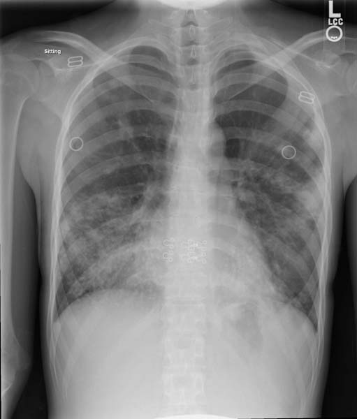
Chest x-ray: Disseminated Tuberculosis
-
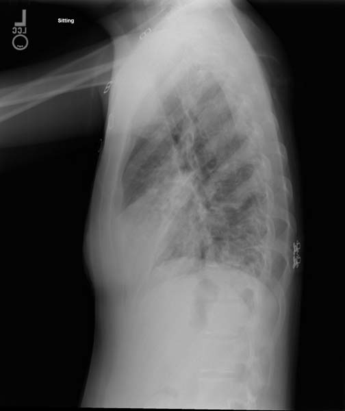
Chest x-ray: Disseminated Tuberculosis


Video showing chest xray in miliary tuberculosis
{{#ev:youtube|xmYE1BlMjno}}
Common findings of miliary tuberculosis on chest x ray
- Fine, pin point approximately 1-2mm in size, discrete, uniform distribution, soft mottlings.
- Commonly found throughout both the lungs.