Testicular vein
Editor-In-Chief: C. Michael Gibson, M.S., M.D. [1]
Overview
The testicular vein (or spermatic vein), the male gonadal vein, carries deoxygenated blood from its corresponding testis to the inferior vena cava or one of its tributaries. It is the male equivalent of the ovarian vein, and is the venous counterpart of the testicular artery.
It a paired vein, with one supplying each testis
- The right testicular vein generally joins the inferior vena cava.
- The left testicular vein, unlike the right, often joins the left renal vein instead of the inferior vena cava.
The veins emerge from the back of the testis, and receive tributaries from the epididymis; they unite and form a convoluted plexus, called the pampiniform plexus, which constitutes the greater mass of the spermatic cord; the vessels composing this plexus are very numerous, and ascend along the cord, in front of the ductus deferens.
Below the subcutaneous inguinal ring they unite to form three or four veins, which pass along the inguinal canal, and, entering the abdomen through the abdominal inguinal ring, coalesce to form two veins, which ascend on the Psoas major, behind the peritoneum, lying one on either side of the internal spermatic artery.
These unite to form a single vein, which opens on the right side into the inferior vena cava, at an acute angle; on the left side into the left renal vein, at a right angle.
The spermatic veins are provided with valves.
The left spermatic vein passes behind the iliac colon, and is thus exposed to pressure from the contents of that part of the bowel.
External links
- Template:SUNYAnatomyLabs - "Posterior Abdominal Wall: Tributaries to the Inferior Vena Cava"
Additional images
-
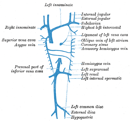
Diagram showing completion of development of the parietal veins.
-
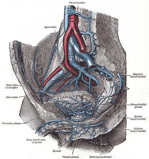
The veins of the right half of the male pelvis.
-

The relations of the viscera and large vessels of the abdomen.
-
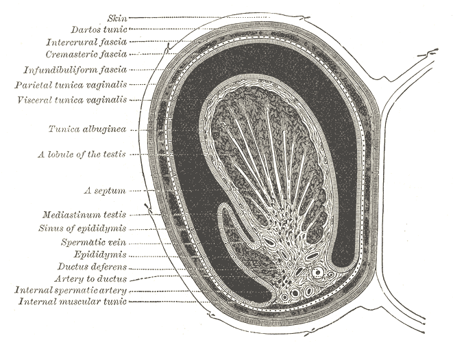
Transverse section through the left side of the scrotum and the left testis.
-
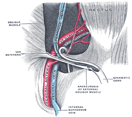
The spermatic cord in the inguinal canal.


