Syphilis physical examination: Difference between revisions
Jump to navigation
Jump to search
| Line 72: | Line 72: | ||
::*Develops 15-20 years after primary infection | ::*Develops 15-20 years after primary infection | ||
::*Clinical presents as [[general paresis]] or [[tabes dorsalis]] with resultant [[ataxia]] | ::*Clinical presents as [[general paresis]] or [[tabes dorsalis]] with resultant [[ataxia]] | ||
::*[[Argyll | ::*[[Argyll Robertson pupil]]: small irregular pupil | ||
===Ophthalmic Examination=== | ===Ophthalmic Examination=== | ||
Revision as of 13:56, 19 December 2012
Editor-In-Chief: C. Michael Gibson, M.S., M.D. [1]
|
Syphilis Microchapters | |
|
Diagnosis | |
|
Treatment | |
|
Case Studies | |
|
Syphilis physical examination On the Web | |
|
American Roentgen Ray Society Images of Syphilis physical examination | |
|
Risk calculators and risk factors for Syphilis physical examination | |
Physical Examination
Primary syphilis: Chancre
- Afebrile
- Chancre:
- Regional lymphadenopathy accompanies primary lesion.
- Onset within a week
- Unilateral or bilateral
- Lymph nodes are firm, painless, non-tender and non-suppurative
- Primary chancre heals spontaneously within 4-6weeks; however, regional lymphadenopathy may persist for longer periods.
Secondary syphilis: Condylomata Lata
- Develops 6-8 weeks after the appearance of primary chancre.
- Cardinal signs include:
- Skin rash: initial macular lesions on the trunk and proximal limbs with progressive generalized papular rash and may cause necrotic ulcers.
- Lymphadenopathy: localized or generalized, firm and non-tender
- Condylomata lata:
- Reddish-brown papular lesions on the intertriginous areas that coalesce and enlarge into large plaques known as condylomata lata
- Lesions usually progress from painful vesicular pattern to erosive lesions with resultant broad, grey-white highly infectious lesions
- Superficial mucosal patches:
-
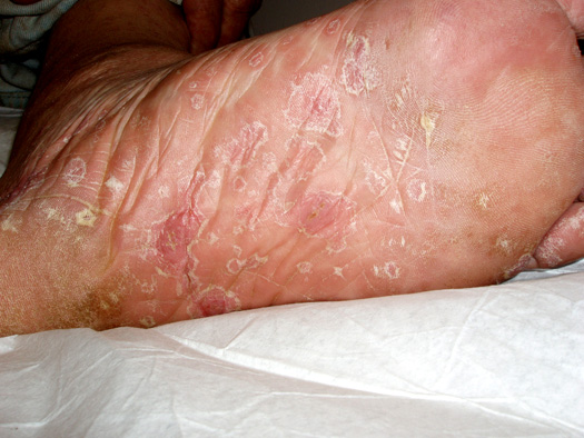
Erruption on Sole of Foot Associated with Secondary Syphilis.
-
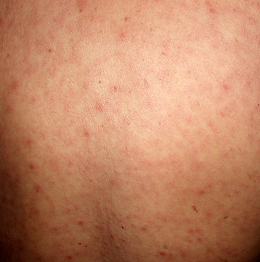
Generalized (Maculo-Papular) Eruption Associated with Secondary Syphilis.
-
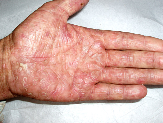
Palmar Erruption Associated with Secondary Syphilis.
-
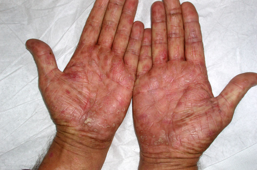
Palmar Erruption Associated with Secondary Syphilis.


Tertiary syphilis: Gumma
- Soft, asymmetric, coalscent granulomatous lesion
- Solitary lesions less than a centimeter in diameter
- Appear almost anywhere in the body including in the skeleton
- Cutaneous gumma: indurated, nodular, papulosquamous to ulcerative lesions with peripheral hyperpigmentation
- Cardiovascular manifestation secondary to aortic dilation with resultant aortic regurgitation:
- Diastolic murmur
- De Musset's sign[1] a bobbing of the head that de Musset first noted in Parisian prostitutes
- Neurological manifestation:
- Asymptomatic meningitis
- Asymptomatic neurosyphilis usually has no signs or symptoms and is diagnosed exclusively with the presence of CSF abnormalities notably pleocytosis, elevated protein, decreased glucose or a positive VDRL test.
- Symptomatic meningitis
- Develops within 6-months to several years of primary infection
- Typical meningitis symptoms present
- Cranial nerve abnormalities may be observed
- Meningovascular syphilis
- Occurs a few months to 10 years (average, 7 years) after the primary infection
- Associated with prodromal symptoms lasting weeks to months before focal deficits are identifiable
- Focal deficits initially are intermittent or progress slowly over a few days
- Clinical present with CNS vascular insufficiency or stroke involving the middle cerebral artery
- Parenchymatous neurosyphilis
- Develops 15-20 years after primary infection
- Clinical presents as general paresis or tabes dorsalis with resultant ataxia
- Argyll Robertson pupil: small irregular pupil
Ophthalmic Examination
- Slit-lamp examination and ophthalmic examination may be helpful to differentiate between acquired and congenital syphilis.
- Presence of interstitial keratitis is suggestive of congenital syphilis with latent infection of unknown duration.
Clinical pearl: Syphilis detecting Handshake
{{#ev:youtube|SAedwyzTMWA}}