Syphilis pathophysiology: Difference between revisions
| Line 12: | Line 12: | ||
==Pathophysiology== | ==Pathophysiology== | ||
===[[Congenital syphilis pathophysiology|Congenital syphilis]]=== | |||
===Acquired syphilis=== | |||
*Incubation period: 3 - 12 weeks | |||
*Spirochete penetrates intact mucous membrane or microscopic dermal abrasions and rapidly enters systemic circulation with the central nervous system being invaded during the early phase of infection. The meninges and blood vessels are initially involved with the brain parenchyma and spinal cord being involved in the later stages of the disease. | |||
*Histopathological hallmark: | |||
:*[[endarteritis]] | |||
:*plasma cell-rich infiltrates reflecting a delayed-type of hypersensitivity to the spirochete | |||
====Primary syphilis==== | |||
''Primary syphilis'' is typically acquired via direct sexual contact with the infectious lesions of a person with syphilis.<ref name=RedBookSyphilis>{{citation | editor=Pickering LK | contribution=Syphilis | title= Red Book | publisher=American Academy of Pediatrics | location= Elk Grove Village, IL | date=2006 | pages=631-644}}</ref> Approximately 10-90 days after the initial exposure (average 21 days), a skin lesion appears at the point of contact, e.g. the [[genitalia]]. This lesion, called a ''[[chancre]]'', is a firm, painless skin ulceration localized at the point of initial exposure to the spirochete, often on the [[penis]], [[vagina]] or [[rectum]]. Rarely, there may be multiple lesions present although typically only one lesion is seen. The [[lesion]] may persist for 4 to 6 weeks and usually heals spontaneously. Local [[lymph node]] swelling can occur. During the initial incubation period, individuals are otherwise [[asymptomatic]]. As a result, many patients do not seek medical care immediately. | |||
Syphilis can ''not'' be contracted through toilet seats, daily activities, hot tubs, or sharing eating utensils or clothing.<ref>{{cite web | last = Centers for Disease Control (CDC) | authorlink = Centers for Disease Control and Prevention | title =STD Facts - Syphilis | publisher = [[Centers for Disease Control]] | date = 05-2004 | url = http://www.cdc.gov/std/syphilis/STDFact-Syphilis.htm }}</ref> | |||
<div align="left"> | |||
<gallery heights="175" widths="175"> | |||
Image:Extragenital syphilitic chancre of the left index finger PHIL 4147 lores.jpg|Primary [[chancre]] of syphilis at the site of infection on the hand | |||
</gallery> | |||
</div> | |||
====Secondary syphilis==== | |||
*CSF: 30% have abnormal findings | |||
==Histopathological Findings== | ==Histopathological Findings== | ||
Revision as of 15:26, 10 February 2012
|
Syphilis Microchapters | |
|
Diagnosis | |
|
Treatment | |
|
Case Studies | |
|
Syphilis pathophysiology On the Web | |
|
American Roentgen Ray Society Images of Syphilis pathophysiology | |
|
Risk calculators and risk factors for Syphilis pathophysiology | |
Editor-In-Chief: C. Michael Gibson, M.S., M.D. [1]
Overview
Casusative organism: Treponema Pallidum
- Small spirochete
- Transmission: requires direct contact with infectious lesion
- Common modes of transmission: vertical transmission, via blood transfusion, via sexual transmission
- Light microscope: Invisible
- Dark field microscope: Distinctive undulating movement seen
Pathophysiology
Congenital syphilis
Acquired syphilis
- Incubation period: 3 - 12 weeks
- Spirochete penetrates intact mucous membrane or microscopic dermal abrasions and rapidly enters systemic circulation with the central nervous system being invaded during the early phase of infection. The meninges and blood vessels are initially involved with the brain parenchyma and spinal cord being involved in the later stages of the disease.
- Histopathological hallmark:
- endarteritis
- plasma cell-rich infiltrates reflecting a delayed-type of hypersensitivity to the spirochete
Primary syphilis
Primary syphilis is typically acquired via direct sexual contact with the infectious lesions of a person with syphilis.[1] Approximately 10-90 days after the initial exposure (average 21 days), a skin lesion appears at the point of contact, e.g. the genitalia. This lesion, called a chancre, is a firm, painless skin ulceration localized at the point of initial exposure to the spirochete, often on the penis, vagina or rectum. Rarely, there may be multiple lesions present although typically only one lesion is seen. The lesion may persist for 4 to 6 weeks and usually heals spontaneously. Local lymph node swelling can occur. During the initial incubation period, individuals are otherwise asymptomatic. As a result, many patients do not seek medical care immediately.
Syphilis can not be contracted through toilet seats, daily activities, hot tubs, or sharing eating utensils or clothing.[2]
-
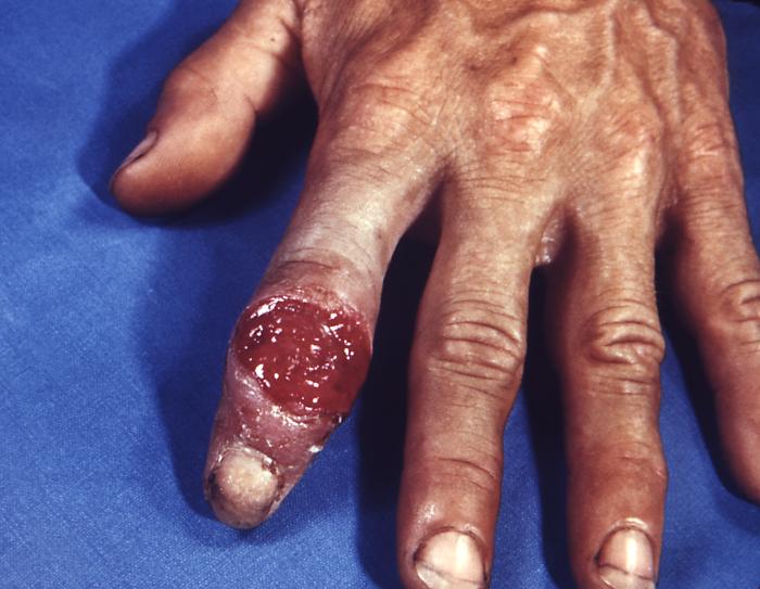
Primary chancre of syphilis at the site of infection on the hand

Secondary syphilis
- CSF: 30% have abnormal findings
Histopathological Findings
Brain: Gumma of syphilis
{{#ev:youtube|Cd60sjchsN8}}
Brain: Paresis (syphilis)
{{#ev:youtube|1Ibu71qHznA}}
References
- ↑ Pickering LK, ed. (2006), "Syphilis", Red Book, Elk Grove Village, IL: American Academy of Pediatrics, pp. 631–644
- ↑ Centers for Disease Control (CDC) (05-2004). "STD Facts - Syphilis". Centers for Disease Control. Check date values in:
|date=(help)