Unused files
Jump to navigation
Jump to search
The following files exist but are not embedded in any page. Please note that other web sites may link to a file with a direct URL, and so may still be listed here despite being in active use.
Showing below up to 500 results in range #51 to #550.
-
 Clip image002.jpg 354 × 227; 11 KB
-
 Timi stemi1.jpg 720 × 540; 35 KB
-
 Timi stemi2.jpg 720 × 540; 28 KB
-
 TIMI NSTEMI1.JPG 720 × 540; 40 KB
-
 TIMI NSTEMI2.JPG 720 × 540; 30 KB
-
 Timi nstemi1.jpg 720 × 540; 40 KB
-
 Timi nstemi2.jpg 720 × 540; 30 KB
-
 Grace score1.JPG 720 × 540; 36 KB
-
 Grace score2.JPG 720 × 540; 42 KB
-
 Grace score3.JPG 720 × 540; 43 KB
-
 Grace score4.JPG 720 × 540; 39 KB
-
 Grace score1.jpg 720 × 540; 36 KB
-
 Grace score2.jpg 720 × 540; 42 KB
-
 Grace score3.jpg 720 × 540; 43 KB
-
 Grace score4.jpg 720 × 540; 39 KB
-
 Popma.jpg 117 × 160; 6 KB
-
 Jeff.Popma.jpg 50 × 75; 26 KB
-
 Jeff Popma.jpg 52 × 75; 34 KB
-
 Abciximab table1.JPG 921 × 465; 55 KB
-
 Abciximab table6.JPG 597 × 459; 33 KB
-
 Lpla art.gif 400 × 347; 65 KB
-
 Cardiomegaly.jpg 957 × 1,021; 57 KB
-
 Cardiomgaly-4.jpg 200 × 180; 6 KB
-
 Aortic valve sa.gif 400 × 347; 52 KB
-
 4 chamber.gif 400 × 347; 72 KB
-
 Apical 2c.gif 400 × 347; 72 KB
-
 Subcostal art.gif 400 × 347; 72 KB
-
 Tamponade.jpg 400 × 320; 19 KB
-
 Foxglove.jpg 300 × 257; 11 KB
-
 Small-bowel-obstruction.jpg 300 × 281; 5 KB
-
 Gingival-hyperlasia.jpg 300 × 279; 5 KB
-
 Measles.jpg 500 × 423; 15 KB
-
 RV-infarct.jpg 300 × 187; 13 KB
-
 Shingles.jpg 500 × 407; 11 KB
-
 Rickets.jpg 91 × 143; 2 KB
-
 Acute-angle-glaucoma.jpg 300 × 198; 6 KB
-
 Spontaneous-pneumothorax.jpg 300 × 282; 13 KB
-
 Diphtheria.jpg 256 × 384; 21 KB
-
 Gonorrhea-epididymitis.jpg 243 × 185; 4 KB
-
 Gonorrhea-gram-stain.jpg 300 × 204; 10 KB
-
 Gonorrhea skin lesion.gif 493 × 307; 77 KB
-
 Ophthalmia neonatorum (gonococcal conjunctivitis).jpg 400 × 307; 28 KB
-
 Crigler-najjar.jpg 200 × 153; 7 KB
-
 Acrodermatitis-enteropathica.jpg 434 × 335; 20 KB
-
 ABI.jpg 402 × 300; 98 KB
-
 Ankle-Brachial Index.jpg 338 × 60; 20 KB
-
 250px-Femoral triangle.gif 250 × 273; 48 KB
-
 Conscious Sedation-definition.jpg 687 × 400; 68 KB
-
 Disease varicella.jpg 125 × 150; 31 KB
-
 Disease varicella2.jpg 125 × 150; 11 KB
-
 5409 thumb.jpg 120 × 80; 2 KB
-
 PHIL 3176 thumb.jpg 100 × 67; 2 KB
-
 PHIL 2290 thumb.jpg 100 × 76; 2 KB
-
 PHIL 2121 thumb.jpg 86 × 80; 2 KB
-
 8153 thumb.jpg 120 × 145; 5 KB
-
 PHIL 2860 thumb.jpg 100 × 71; 2 KB
-
 6114 thumb.jpg 120 × 79; 3 KB
-
 6545 thumb.jpg 120 × 77; 3 KB
-
 PHIL 3719 thumb.jpg 100 × 66; 2 KB
-
 Bidmc.gif 57 × 63; 2 KB
-
 Hmsshield.gif 57 × 57; 3 KB
-
 Shapring ribbon.jpg 536 × 80; 45 KB
-
 Spring coil outer only.jpg 697 × 57; 36 KB
-
 Spring coil tip only.jpg 676 × 61; 27 KB
-
 Covers.jpg 723 × 62; 30 KB
-
 Relationship of lubricity to tactile feedback.jpg 852 × 435; 92 KB
-
 Joint wire bends.jpg 452 × 121; 21 KB
-
 Small-pills.jpg 80 × 53; 2 KB
-
 Wikidoc-living-textbook.jpg 154 × 147; 8 KB
-
 Wikidoc-living-textbook-small.jpg 80 × 76; 2 KB
-
 RBBB.LAFB.long PR.jpg 1,642 × 1,032; 161 KB
-
 ARDS.JPG 466 × 404; 9 KB
-
 180px-Poumon artificiel.jpg 180 × 128; 5 KB
-
 Svc-syndrome.jpg 289 × 379; 20 KB
-
 Barrel chest.jpg 1,000 × 750; 61 KB
-
 Christine Bjerke.jpg 63 × 75; 13 KB
-
 TING.jpg 53 × 75; 12 KB
-
 Disease yellow fever.jpg 125 × 150; 16 KB
-
 Trichuris eggC.jpg 200 × 200; 5 KB
-
 Trichuris eggD.jpg 200 × 200; 5 KB
-
 KathyKastan.jpg 50 × 75; 29 KB
-
 Map Africa--Yellow Fever.jpg 153 × 200; 25 KB
-
 Map South America--Yellow Fever.jpg 152 × 200; 22 KB
-
 Xenopsylla Cheopis.jpg 120 × 80; 14 KB
-
 World98 sm.gif 224 × 150; 7 KB
-
 Map WNV 807 07 a.jpg 363 × 220; 30 KB
-
 DSCN1357.JPG 2,816 × 2,112; 869 KB
-
 TimHenry.jpg 1,660 × 1,664; 201 KB
-
 TimHenry2.jpg 500 × 501; 80 KB
-
 TimHenry3.jpg 75 × 75; 18 KB
-
 TimHenry4.jpg 125 × 125; 23 KB
-
 Buerger's Disease.JPG 250 × 201; 23 KB
-
 Hepatitis c-risk.JPG 875 × 1,354; 69 KB
-
 Hepatitis D virus.gif 550 × 358; 30 KB
-
 Hepatits E from CDC.jpg 444 × 342; 27 KB
-
 Hepatitis E distribution from CDC.GIF 406 × 305; 9 KB
-
 JCB portrait.jpg 56 × 75; 16 KB
-
 Non-cholera Vibrio infections.jpg 809 × 506; 59 KB
-
 Smallpox.gif 300 × 355; 28 KB
-
 Smallpox virus particle.jpg 700 × 564; 59 KB
-
 Ulcer.jpg 268 × 515; 27 KB
-
 Wikidoc-on-your-ipod.jpg 359 × 495; 27 KB
-
 Typhus fevers.jpg 570 × 233; 48 KB
-
 Wikidoc-on-your-ipod-2.jpg 357 × 389; 20 KB
-
 Typhus fever table.jpg 570 × 289; 55 KB
-
 Treatment of Patients With Tularemia.jpg 590 × 502; 70 KB
-
 Tularemia.jpg 594 × 504; 70 KB
-
 Treatment tuberculosis 1.jpg 662 × 445; 45 KB
-
 Drug regimens for tuberculosis.jpg 708 × 416; 111 KB
-
 Kaposi's sarcoma.jpg 700 × 583; 56 KB
-
 African trypanosomiasis 2 and 3.jpg 349 × 402; 36 KB
-
 African trypanosomiasis 6.jpg 384 × 274; 25 KB
-
 Kissing bug.jpg 163 × 117; 16 KB
-
 Jauch2.jpg 52 × 75; 21 KB
-
 Pneumocystis jiroveci.jpg 700 × 472; 45 KB
-
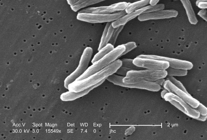 Tuberculosis SEM.jpg 700 × 475; 49 KB
-
 Toxoplasmosi AIDS.jpg 700 × 472; 41 KB
-
 Aortic stenosis.jpg 700 × 466; 88 KB
-
 Acanthocytosis.jpg 700 × 475; 58 KB
-
 P acnes.jpg 521 × 500; 49 KB
-
 Actinomycosis 2.jpg 700 × 596; 29 KB
-
 LASMA still4.jpg 512 × 512; 148 KB
-
 Figure 1 copy.jpg 466 × 278; 40 KB
-
 Table 2.jpg 465 × 344; 64 KB
-
 Figure 2.jpg 462 × 310; 38 KB
-
 Table3.jpg 458 × 519; 72 KB
-
 Figure 3.jpg 463 × 334; 39 KB
-
 Fig3.jpg 457 × 334; 39 KB
-
 Figure 4.jpg 460 × 334; 38 KB
-
 Figure 5.jpg 457 × 289; 36 KB
-
 Figure 6.jpg 459 × 440; 78 KB
-
 Life cycle of Trichinella spp.jpg 449 × 547; 68 KB
-
 Trichinella A and B.jpg 420 × 218; 26 KB
-
 Trichinella C and D.jpg 343 × 192; 25 KB
-
 Syphilis-Reported cases by stage of infection.jpg 996 × 466; 61 KB
-
 Figure 2--Primary and secondary syphilis — Rates by region.jpg 1,004 × 491; 79 KB
-
 Primary and secondary syphilis.jpg 1,007 × 533; 88 KB
-
 Primary and secondary syphilis — Rates by county.jpg 1,005 × 605; 122 KB
-
 Prim and second syphilis — Rates by county.jpg 1,010 × 584; 113 KB
-
 Primary and secondary syphilis — Rates Total and by sex.jpg 1,005 × 465; 65 KB
-
 Pink-eye.jpg 150 × 75; 3 KB
-
 Toxoplasmosis.jpg 207 × 362; 20 KB
-
 Cocreation-small-1.jpg 300 × 77; 8 KB
-
 Cocreate-small-2.jpg 298 × 124; 13 KB
-
 Cocreate-3.jpg 298 × 124; 13 KB
-
 Treatment.jpg 519 × 290; 48 KB
-
 TBRF by state.jpg 507 × 392; 14 KB
-
 Rubella.jpg 391 × 309; 33 KB
-
 Rubella virus.jpg 131 × 158; 22 KB
-
 Selected vaccine-preventable diseases, United States.jpg 460 × 320; 49 KB
-
 Tetanus.jpg 391 × 156; 26 KB
-
 Tetanus--United States 1947-2004.jpg 369 × 236; 24 KB
-
 Tetanus--United States 1980-2004.jpg 368 × 236; 23 KB
-
 Tetanus--United States Age Distribution.jpg 368 × 235; 25 KB
-
 Tetanus--Injuries and Conditions.jpg 367 × 245; 26 KB
-
 Tuberculosis x-ray.jpg 704 × 547; 42 KB
-
 Diphyllobothrium latum egg.jpg 425 × 242; 27 KB
-
 Diphyllobothrium latum 2.jpg 498 × 241; 32 KB
-
 Diphyllobothrium latum proglottids.jpg 518 × 245; 24 KB
-
 Diphyllobothrium latum proglottids 2.jpg 224 × 245; 34 KB
-
 Kids in pool.gif 141 × 197; 19 KB
-
 Early-Onset Disease Incidence.jpg 1,377 × 1,053; 89 KB
-
 Graph of Early-Onset Disease Incidence.jpg 367 × 281; 32 KB
-
 Thumb1.gif 440 × 330; 27 KB
-
 Thumb4.gif 440 × 330; 29 KB
-
 Thumb5.gif 440 × 330; 32 KB
-
 Box1.gif 694 × 830; 91 KB
-
 Incidence.gif 334 × 336; 15 KB
-
 Cases by state.jpg 600 × 364; 44 KB
-
 Cases by year.jpg 535 × 349; 21 KB
-
 PainReliever.gif 165 × 250; 14 KB
-
 Amblyomma-americanum.jpg 575 × 546; 65 KB
-
 Feline sporotrichosis 4.jpg 300 × 233; 14 KB
-
 News.jpg 175 × 131; 10 KB
-
 Hanta trapgloves21.gif 200 × 117; 19 KB
-
 53 ov.jpg 300 × 194; 12 KB
-
 Schmfig.gif 600 × 449; 84 KB
-
 Episl2.gif 500 × 333; 40 KB
-
 Episl4.gif 500 × 333; 29 KB
-
 Episl5.gif 500 × 333; 40 KB
-
 Disease shingles.jpg 125 × 150; 34 KB
-
 Disease shingles2.jpg 125 × 150; 13 KB
-
 Table.gif 483 × 362; 13 KB
-
 Box.gif 474 × 444; 28 KB
-
 M214a1t.gif 473 × 741; 25 KB
-
 M214a1f.gif 689 × 364; 14 KB
-
 Coronavirus.JPG 174 × 125; 7 KB
-
 500px-Aisdiag.png 500 × 381; 38 KB
-
 164px-Chromosome X.svg.png 164 × 450; 16 KB
-
 Chromosome X.svg.png 164 × 450; 16 KB
-
 Gruntzig.jpg 164 × 200; 33 KB
-
 Willem.jpg 460 × 600; 38 KB
-
 Pale-lad-with-dsa-small.gif 110 × 113; 684 KB
-

-
 Test slides.ppt ; 132 KB
-
 Figure 1.png 362 × 275; 10 KB
-
 Correct timing.JPG 335 × 240; 12 KB
-
 Early inflation.JPG 422 × 256; 11 KB
-
 Early deflation.JPG 421 × 254; 13 KB
-
 Late deflation.JPG 423 × 259; 12 KB
-
 Late inflation.JPG 423 × 250; 12 KB
-
MOV00019.swf ; 1.18 MB
-
 Preoperative Clearance.jpg 640 × 799; 60 KB
-
 Follicular adenoma of the thyroid.jpg 261 × 400; 23 KB
-
 800px-Test biosynth 17BHSD3.jpg 800 × 556; 38 KB
-
 Cretin Child (1).jpg 800 × 535; 108 KB
-
 560px-DHEA1.svg.png 560 × 225; 16 KB
-
 DHEA1.svg.png 560 × 225; 16 KB
-
 Hood and glans labeled.jpg 317 × 129; 34 KB
-
 Autorecessive.svg.png 512 × 599; 52 KB
-
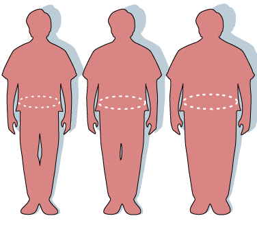
 Central Obesity 008.jpg 606 × 600; 38 KB
-
 Brain met.jpg 511 × 599; 39 KB
-
 Glioblastoma - MR coronal with contrast.jpg 617 × 600; 57 KB
-
 Glioblastoma - MR sagittal with contrast.jpg 612 × 599; 59 KB
-
 Glioblastoma (1).jpg 600 × 452; 94 KB
-
 Cholecalciferol.jpg 560 × 599; 10 KB
-
 Ergocalciferol.svg.jpg 560 × 599; 11 KB
-
 InsulinHexamer.jpg 640 × 434; 41 KB
-
 BCtimeline.jpg 800 × 481; 71 KB
-
 Stomach diagram.svg.png 255 × 255; 22 KB
-
 Linitis plastica 2.jpg 631 × 580; 77 KB
-
 Prostatehistopath.jpg 800 × 583; 165 KB
-
 Linacprostate.jpg 324 × 385; 19 KB
-
 Hugginsprostate.jpg 665 × 479; 217 KB
-
 Malignant melanoma.jpg 320 × 240; 154 KB
-
 WB032021.JPG 800 × 532; 73 KB
-
 Malignant melanoma (1) at thigh Case 01.jpg 600 × 452; 72 KB
-
 Malignant melanoma (4) at thigh Case 01.jpg 600 × 452; 72 KB
-
 Superficial spreading melanoma 1 060619.jpg 220 × 165; 41 KB
-
 MMatthigh.jpg 600 × 452; 84 KB
-
 Mid esophageal mass.jpg 282 × 523; 24 KB
-
 Esophagael stent.jpg 715 × 599; 61 KB
-
 Lymphoma microarray.jpg 800 × 584; 178 KB
-
 Renal cell ca.jpg 431 × 600; 70 KB
-
 RCC.jpg 512 × 512; 55 KB
-
 Bonetumor.jpg 472 × 354; 73 KB
-
 Osteochondroma right scapula.jpg 800 × 406; 44 KB
-
 Terry fox running.jpg 250 × 356; 16 KB
-
 Canine osteosarcoma.JPG 800 × 600; 19 KB
-
 Basaliom1.jpg 600 × 430; 146 KB
-
 Basaliom2.jpg 600 × 586; 185 KB
-
 Basaliom3.jpg 600 × 487; 166 KB
-
 Oxytocin.jpg 493 × 413; 127 KB
-
 Rickewrist1.jpg 640 × 480; 23 KB
-
 Life cycle schistosomiasis.jpg 532 × 476; 77 KB
-
 Schistosoma japonicum.jpg 421 × 239; 31 KB
-
 Schistosoma j.jpg 423 × 242; 32 KB
-
 Map s.jpg 557 × 456; 63 KB
-
 Map of distribution.jpg 555 × 429; 58 KB
-
 Pic.jpg 623 × 664; 71 KB
-
 Scarlet fever.jpg 144 × 213; 19 KB
-
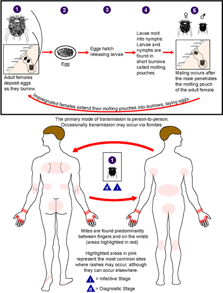 Scabies LifeCycle.gif 435 × 570; 38 KB
-
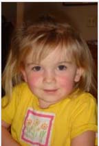
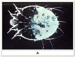 Scabei mite.jpg 307 × 236; 33 KB
-
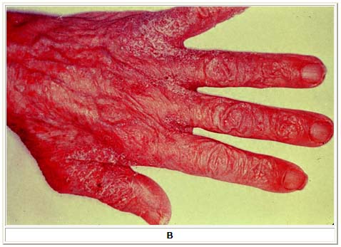 Scabies lesions.jpg 478 × 347; 49 KB
-
 Horse spleen laparoscopic.jpg 375 × 288; 23 KB
-
 Obesity-waist circumference.PNG 376 × 331; 18 KB
-
 Saladeespera12.jpg 640 × 480; 39 KB
-
 MeSH-example.svg.png 256 × 256; 14 KB
-
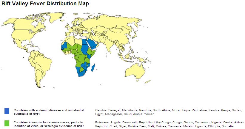 RVF map.jpg 821 × 434; 63 KB
-
 A. lumbricoides female worm.jpg 270 × 237; 27 KB
-
 Regional.jpg 563 × 410; 56 KB
-
 FEET2.gif 252 × 198; 11 KB
-
 Ricketts.gif 123 × 200; 8 KB
-
 Cells.jpg 297 × 278; 6 KB
-
 Epidem1.gif 419 × 207; 9 KB
-
 EpiGraph1.gif 440 × 265; 10 KB
-
 EpiGraph2.gif 450 × 289; 13 KB
-
 RMSFMAP.gif 410 × 310; 22 KB
-
 Table.jpg 546 × 411; 89 KB
-
 Arm.jpg 302 × 292; 8 KB
-
 Rick-IHC.jpg 603 × 404; 21 KB
-
 Bullet.gif 389 × 164; 24 KB
-
 30th-anniversary-banner.jpg 841 × 200; 105 KB
-
Template for heartscape.ppt ; 105 KB
-
Links to Online Resources.doc ; 75 KB
-
 Map rabies.jpg 449 × 297; 57 KB
-
 Map US rabies.jpg 500 × 331; 61 KB
-
 Lambs2.jpg 215 × 238; 32 KB
-
 Crab lice.jpg 700 × 519; 54 KB
-
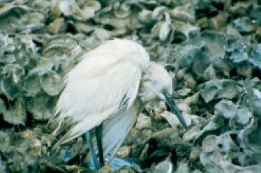 Blue heron chlamydiosis.jpg 374 × 248; 27 KB
-
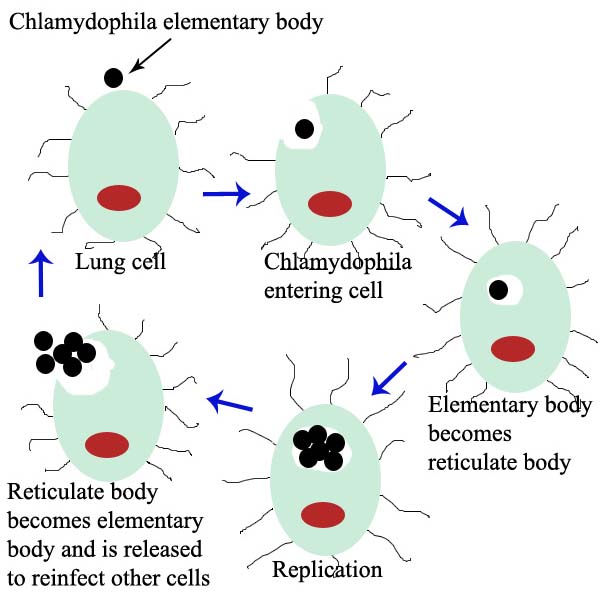 Chlamydophila body.jpg 604 × 594; 67 KB
-
 Seniors.jpg 200 × 132; 8 KB
-
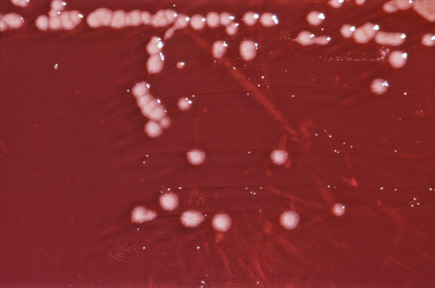 Pseudomonas a..jpg 1,817 × 1,206; 1.18 MB
-
 Legionellosis table.jpg 786 × 581; 114 KB
-
 CMR pics.jpg 899 × 718; 80 KB
-
 Shax.jpg 416 × 466; 23 KB
-
 Polio USA, 1950-2004.jpg 329 × 248; 23 KB
-
 Polio USA, 1980-2004.jpg 329 × 252; 27 KB
-
 Polio lores134.jpg 1,024 × 1,536; 256 KB
-
 Polio lores.jpg 302 × 450; 38 KB
-
 Polio pic.jpg 362 × 540; 44 KB
-
 Poliovirus.jpg 438 × 604; 101 KB
-
 Poliovirus Myotonic dystrophic.jpg 875 × 590; 99 KB
-
 Polio spinal diagram.jpg 806 × 595; 70 KB
-
 Brain bulbar region.jpg 319 × 314; 31 KB
-
 A gambiae.jpg 200 × 130; 6 KB
-
 Plasmodium f.jpg 120 × 90; 2 KB
-
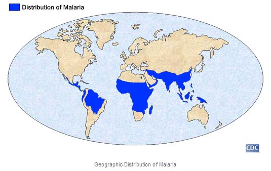 Malaria map.jpg 535 × 338; 44 KB
-
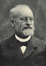 Laveran.jpg 150 × 214; 7 KB
-
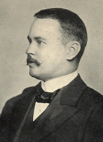 Ronald Ross.jpg 150 × 207; 8 KB
-
 Little patient.jpg 240 × 161; 15 KB
-
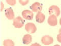
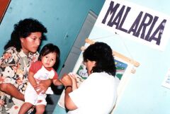
 Malaria patient.jpg 240 × 161; 15 KB
-
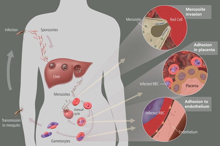 Malaria cycle.jpg 700 × 467; 38 KB
-
 Beef cattle grazing usda.gif 250 × 164; 25 KB
-
 BSE chart.gif 960 × 720; 13 KB
-
 Aphis usda.jpg 752 × 599; 114 KB
-
 Gorgeous Cow.jpg 900 × 838; 187 KB
-
 BSE table.jpg 486 × 388; 53 KB
-
 Cow with BSE.jpg 750 × 600; 94 KB
-
 CJD--pathologic and clinical.jpg 559 × 536; 116 KB
-
 Classic CJD patient--tissue.jpg 511 × 386; 50 KB
-
Wiki perfusion.swf ; 4.77 MB
-
 VCJD percent distribution.jpg 508 × 370; 72 KB
-
 PediculusHumanus.png 651 × 600; 79 KB
-
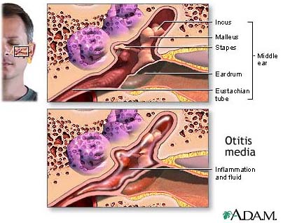 Otitis media.jpg 404 × 318; 55 KB
-
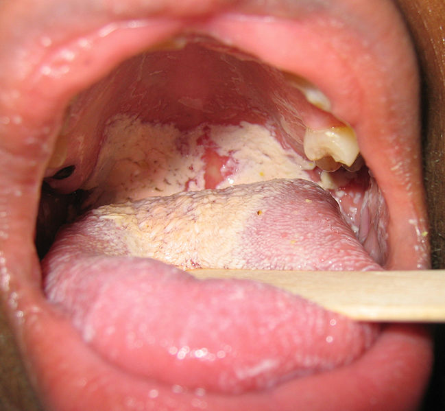 Oralcandi.jpg 649 × 599; 60 KB
-
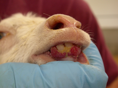 Orf goat kid.jpg 400 × 300; 86 KB
-
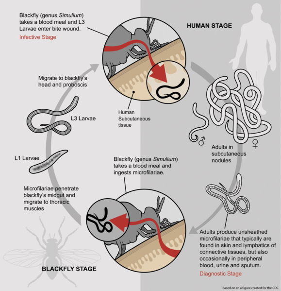 Life Cycle of Onchocerca.jpg 579 × 599; 56 KB
-
 Onchocerca.jpg 800 × 538; 11 KB
-
 Coldagglutinin.svg.png 797 × 580; 259 KB
-
 Hairy cell leukemia.jpg 433 × 295; 103 KB
-
 Dexamethasone-2D-skeletal.png 1,100 × 846; 45 KB
-
 Anthrax - inhalational.jpg 343 × 284; 88 KB
-
 Powell-anthrax-vial.jpg 410 × 285; 16 KB
-
 Antibody.svg.png 255 × 360; 12 KB
-
 Template-info.svg.png 50 × 26; 3 KB
-
 Spina bifida drawing.gif 190 × 243; 8 KB
-
 IMG 0746a.jpg 500 × 375; 16 KB
-
 Diphtheria map.png 800 × 370; 88 KB
-
 Neisseria gonorrhoeae.jpg 700 × 456; 25 KB
-
 Naegleria fowleri trophozoite.jpg 217 × 242; 23 KB
-
 Naegleria fowleri.jpg 800 × 530; 75 KB
-
 People mumps.jpg 125 × 150; 31 KB
-
 PHIL 1874.jpg 296 × 294; 71 KB
-
 Epstein Barr Virus virions.jpg 800 × 570; 113 KB
-
 Parainfluenza virus.jpg 652 × 500; 62 KB
-
 AoDissekt scheme StanfordB.png 498 × 599; 252 KB
-
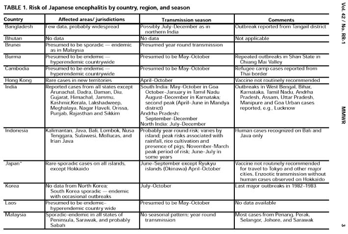 Table japanese encephalitis.jpg 710 × 477; 107 KB
-
 Table japanese encephalitis 2.jpg 710 × 481; 114 KB
-
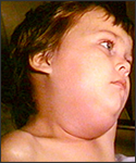
 Map of Japanese encephalitis.jpg 600 × 541; 60 KB
-
 Mastomys.gif 175 × 99; 15 KB
-
 Lassa virus.jpg 800 × 589; 89 KB
-
 Histoplasmosis skin lesion .jpg 700 × 475; 32 KB
-
 Femoral 1.png 497 × 456; 142 KB
-
 Haemophilus i..jpg 800 × 539; 86 KB
-
 Leprosy.jpg 440 × 599; 50 KB
-
 Leprae.jpg 800 × 537; 141 KB
-
 Mycobacterium leprae.jpg 489 × 500; 55 KB
-
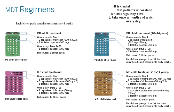 MDTRegimens.jpg 674 × 435; 161 KB
-
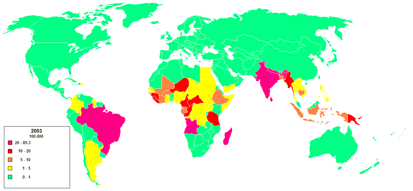 Lepra 2003.png 1,357 × 628; 20 KB
-
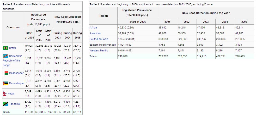 Globlal leprosy.jpg 1,066 × 485; 100 KB
-
 Leprosy cutaneous lesions.jpg 800 × 534; 62 KB
-
 Dracunculiasis.gif 593 × 435; 27 KB
-
 Dracunculiasis feet.jpg 578 × 227; 33 KB
-
 Guinea worm.jpg 250 × 165; 14 KB
-
 ASD diagram.jpg 217 × 177; 17 KB
-
 ASD map.jpg 308 × 277; 4 KB
-
 Padlock.svg.png 40 × 40; 2 KB
-
 C Michael Gibson.jpg 200 × 272; 29 KB
-
 Sinus venosus defect.jpg 200 × 186; 9 KB
-
 Cha Yong-Mei 01F.jpg 63 × 75; 26 KB
-
 NFPA 704.svg.png 600 × 600; 8 KB
-
 Chvostek Sign.PNG 256 × 308; 11 KB
-
 Chvostek Appearance.PNG 183 × 216; 43 KB
-
 Summary.png 476 × 481; 32 KB
-
 Common lipids.png 790 × 599; 101 KB
-
 Fig6T1T2.jpg 919 × 275; 18 KB
-
 Fig8pulseseq.jpg 1,262 × 525; 36 KB
-
 Flag of WHO.svg 800 × 533; 59 KB
-
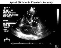 Apical 4 chamber Ebstein.jpg 200 × 157; 7 KB
-
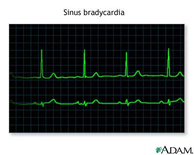 Bradycardia.jpg 400 × 320; 16 KB
-
 UterineCancerTumor.jpg 150 × 113; 4 KB
-
 Thompson's Gazelle.jpg 187 × 156; 14 KB
-
 355px-Human-gender-neutral.png 355 × 599; 99 KB
-
 Human-gender-neutral.png 355 × 599; 99 KB
-
 Eptifibatide.svg 800 × 541; 28 KB
-
 1bda.png 640 × 434; 83 KB
-
 Accuracy and precision.png 520 × 280; 12 KB
-
 Binomial distribution cdf.png 561 × 420; 5 KB
-
 AngiotensinCE-1O8A.png 424 × 358; 89 KB
-
 12leadECG.jpg 1,040 × 407; 73 KB
-
 Heart attack diagram.png 522 × 372; 27 KB
-
 ECG 001.jpg 800 × 431; 67 KB
-
 Myocardial infarct emmolition phase histopathology.jpg 790 × 599; 289 KB
-
 AAA-101.jpg 559 × 488; 55 KB
-
 Gnome globe current event svg.png 600 × 600; 139 KB
-
 Coarctation.png 480 × 599; 91 KB
-
 Cooley-Liotta-Mr HaskellKarp.jpg 250 × 172; 60 KB
-
 Endo dysfunction Athero.png 546 × 600; 310 KB
-
 Surgeon operating Fitzsimons Army Medical Center circa 1990.jpg 402 × 599; 69 KB
-
 Surgeon operating, Fitzsimons Army Medical Center, circa 1990.jpg 402 × 599; 69 KB
-
 Heart-thorax-gray.gif 422 × 400; 24 KB
-
 Sports-dress-codes.jpg 475 × 396; 62 KB
-
 Cardiac vessels.png 960 × 1,440; 73 KB
-
 Digitoxin svg.png 519 × 278; 10 KB
-
 Pacemaker GuidantMeridianSR.jpg 549 × 600; 66 KB
-
 Digitoxin.png 519 × 278; 10 KB
-
 Heartchambers.jpg 400 × 320; 27 KB
-
 Hemangiosarcoma.jpg 800 × 600; 102 KB
-
 Iron metabolism svg.png 500 × 250; 17 KB
-
 Holter.jpg 120 × 120; 7 KB
-
 Cholesterol svg.png 440 × 296; 14 KB
-
 Cardiovasc Ultrasound LVNC 1.jpg 400 × 278; 26 KB
-
 Cardiovasc Ultrasound LVNC 2.jpg 400 × 575; 45 KB
-
 Cardiovasc Ultrasound LVNC 3.jpg 400 × 240; 21 KB
-
 Cardiovasc Ultrasound LVNC 4.jpg 400 × 235; 19 KB
-
 Statin.jpg 218 × 310; 12 KB
-
 Tetralogy of Fallot svg.png 614 × 441; 109 KB
-
 Glanzstreifen.jpg 750 × 600; 105 KB
-
 Heart cross section.jpg 400 × 320; 19 KB
-
 St Jude Medical pacemaker with ruler.jpg 566 × 600; 74 KB
-
 Common Flutter.png 716 × 335; 194 KB
-
 EKG VF.jpg 800 × 196; 49 KB
-
 ICD.jpg 640 × 480; 176 KB
-
 SV Tachycardia marked.jpg 800 × 207; 62 KB
-
 Ottawa Heart Institute-.jpg 800 × 600; 103 KB
-
 Flag of Ottawa, Ontario.svg.png 432 × 216; 15 KB
-
 Tirofiban.svg 800 × 238; 10 KB
-
 Zbigniew Religa.jpg 115 × 160; 8 KB
-
 Right Ventricular hypertrophy svg.png 619 × 451; 101 KB
-
 Sodium-nitroprusside-2D.png 800 × 561; 30 KB
-
 Cyanosis.jpg 400 × 320; 9 KB
-
 Blood culture negative endocarditis.jpg 500 × 326; 33 KB
-
 CT pericardial effusion.jpg 726 × 600; 52 KB
-
 Assessory meningeal artery.png 600 × 540; 57 KB
-
 HOCM.JPG 389 × 397; 13 KB
-
 HOCM ECG.png 400 × 344; 72 KB
-
 HOCM LVH.jpg 320 × 240; 23 KB
-
 Ventricular assist device.jpg 300 × 450; 30 KB
-
 Illu quiz lung04.jpg 270 × 350; 42 KB
-
 Illu quiz lung02.jpg 270 × 350; 42 KB
-
 Aortic dissection (1) Victoria blue-HE.jpg 500 × 376; 87 KB
-
 VF.jpg 900 × 221; 57 KB
-
 Vt.jpg 900 × 342; 40 KB
-
 Heart numlabels.png 957 × 965; 237 KB
-
 Normalandfallot.jpg 438 × 290; 41 KB
-
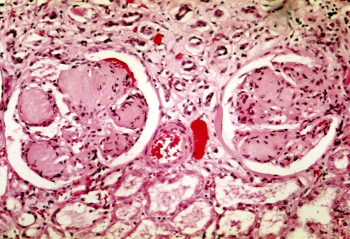 Nodular glomerulosclerosis.jpg 700 × 479; 84 KB
-
 Blood Coagulation and Protein C Pathways.jpg 787 × 584; 65 KB
-
 Protein C anticoagulant.jpg 624 × 348; 22 KB
-
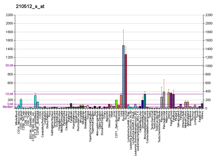 PBB GE VEGFA 210512 s at fs.png 732 × 530; 11 KB
-
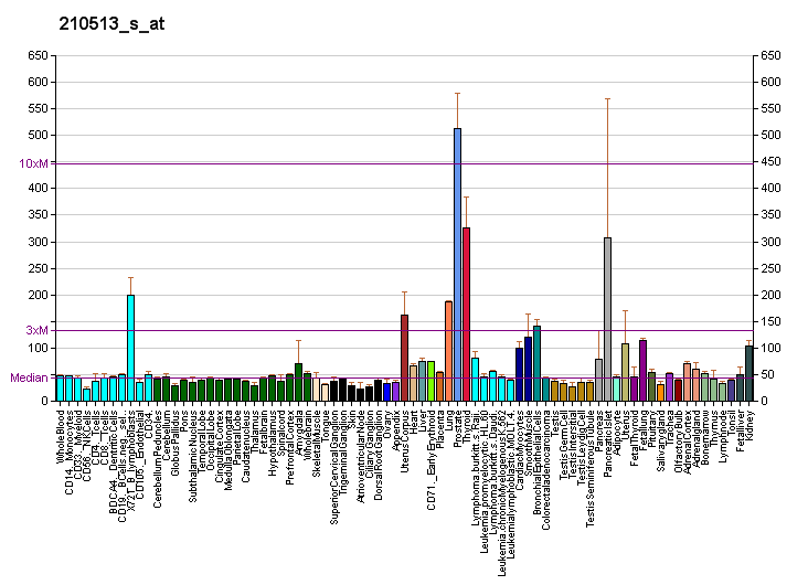 PBB GE VEGFA 210513 s at fs.png 732 × 530; 12 KB
-
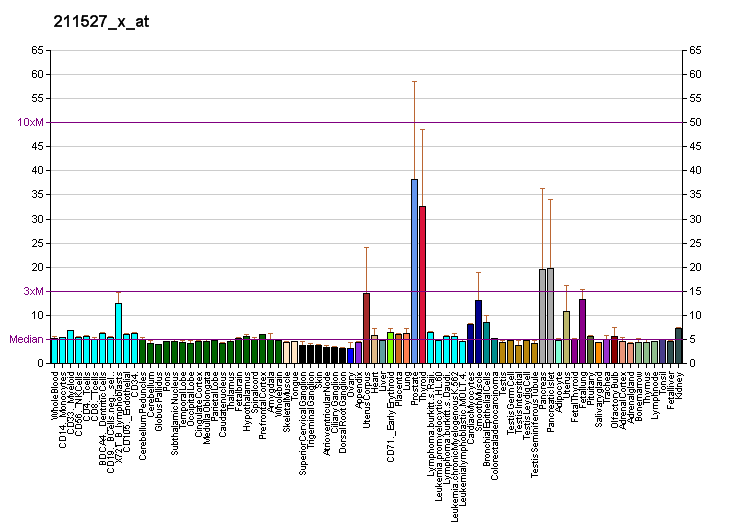 PBB GE VEGFA 211527 x at fs.png 732 × 530; 11 KB
-
 Sagital aaa.jpg 519 × 918; 142 KB
-
 Dog heart 2.jpg 1,787 × 2,427; 504 KB
-
 Celiac a branches stomach in situ.gif 650 × 669; 123 KB
-
 Gray1237 svg.png 428 × 599; 113 KB
-
 External carotid a.gif 600 × 647; 138 KB
-
 Femoral artery and branches.gif 327 × 600; 33 KB
-
 Gray1086-liver.png 675 × 465; 81 KB
-
 Schematic diagram of the human eye en svg.png 508 × 516; 85 KB
-
 Benzoic-acid.svg 153 × 89; 2 KB
-
 Benzoic-acid-3D-vdW.png 755 × 600; 109 KB
-
 Parathyroidglands.png 414 × 500; 52 KB
-
 Internaliliac.png 557 × 600; 462 KB
-
 Inferior mesenteric a.gif 535 × 650; 97 KB
-
 Midrectal.png 700 × 754; 186 KB
-
 Popliteal artery.png 461 × 1,000; 172 KB
-
 Posterior auricular.png 700 × 647; 186 KB
-
 Superior mesenteric a.gif 500 × 609; 93 KB
-
 Superior thyroid.png 629 × 647; 182 KB
-
 Axillary branches.png 800 × 600; 140 KB
-
 Category-menu-system.jpg 173 × 470; 33 KB
-
 AS and coronaries.jpg 300 × 200; 18 KB
-
 Box2.jpg 325 × 55; 2 KB
-
 Aortic stenosis rheumatic gross path 20G0014 lores rotated.jpg 700 × 466; 68 KB
-
 Inferiorvesical.png 700 × 754; 186 KB
-
 Cavernous sinus.png 500 × 601; 163 KB
-
 Internaliliacv.png 500 × 580; 76 KB
-
 Portal vein and tributaries.gif 491 × 750; 287 KB
-
 ABO blood type svg.png 796 × 555; 92 KB
-
 ABO blood group diagram svg.png 795 × 600; 139 KB
-
 Codominant.jpg 348 × 396; 28 KB
-
 RhO+.jpg 320 × 240; 16 KB
-
 RhB-.jpg 320 × 240; 16 KB
-
 Band 3 Atomic microscope.jpg 554 × 387; 32 KB
-
 Blood Donation 12-07-06 1.jpg 1,632 × 1,224; 396 KB
-
 Blood Donation 12-07-06 2.jpg 1,632 × 1,224; 355 KB
-
 Blood donation bruise.jpg 1,000 × 751; 100 KB
-
 Blood Compatibility svg.png 559 × 539; 71 KB
-
 Hemorrhoids2.jpg 589 × 456; 83 KB
-
 Hemorrhoids.jpg 714 × 590; 120 KB
-
 Diagram.png 624 × 567; 17 KB
-
 Head deep facial trigeminal.jpg 1,205 × 1,424; 1.18 MB
-
 Abducens nerve1.png 615 × 459; 198 KB
-
 Cranial nerve VII svg.png 424 × 532; 129 KB
-
 Ramizygomaticinervifacialis.png 512 × 600; 444 KB
-
 Ramusmarginalismandibularisnervifacialis.png 512 × 600; 441 KB
-
 Wavefront-for-web.gif 65 × 65; 15 KB
-
 Plaquerupture.jpg 741 × 475; 81 KB
-
 PBB Protein F10 image.jpg 500 × 500; 42 KB
-
 LL picture.jpg 554 × 384; 27 KB
-
 Ciliary ganglion.png 606 × 396; 219 KB
-
 Tsetse fly.png 306 × 257; 17 KB
-
 JeanChrétien.jpg 219 × 217; 12 KB
-
 Bells.jpg 500 × 374; 157 KB
-
 Aerosol from Sneeze.jpg 800 × 533; 97 KB
-
 PHIL 3183 lores.jpg 700 × 459; 27 KB
-
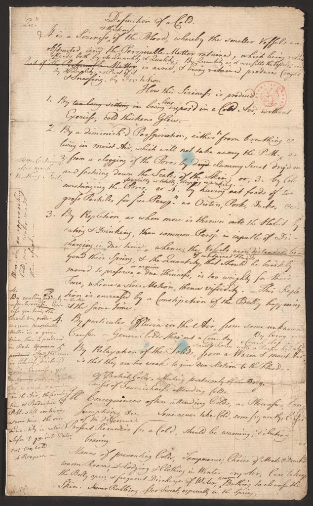 Definition of a Cold by Benjamin Franklin Page 1.jpg 634 × 1,024; 152 KB
-
 Chalazion25pc.jpg 160 × 120; 30 KB


.jpg)


.jpg)


.jpg)


_at_thigh_Case_01.jpg)
_at_thigh_Case_01.jpg)


_Victoria_blue-HE.jpg)


