Sexcord/ stromal ovarian tumors other imaging findings: Difference between revisions
No edit summary |
|||
| (7 intermediate revisions by the same user not shown) | |||
| Line 2: | Line 2: | ||
{{Sexcord/ stromal ovarian tumors}} | {{Sexcord/ stromal ovarian tumors}} | ||
{{CMG}}; {{AE}} | {{CMG}}; {{AE}};{{M.N}} | ||
==Overview== | ==Overview== | ||
PET-CT may be helpful in the diagnosis of sexcord/ stromal ovarian tumors | [[PET scan|PET]]-CT may be helpful in the [[diagnosis]] of sexcord/ stromal ovarian tumors. On [[fluorodeoxyglucose]]-[[positron emission tomography]]/[[computed tomography]] [[malignant]] ovarian tumors generally have intense uptake, whereas [[tumors]] with a small [[solid]] content show decreased uptake. | ||
==Other Imaging Findings== | ==Other Imaging Findings== | ||
PET-CT may be helpful in the diagnosis sexcord/ stromal ovarian tumors <ref name="pmid25886261">{{cite journal |vauthors=Qian Q, You Y, Yang J, Cao D, Zhu Z, Wu M, Chen J, Lang J, Shen K |title=Management and prognosis of patients with ovarian sex cord tumor with annular tubules: a retrospective study |journal=BMC Cancer |volume=15 |issue= |pages=270 |date=April 2015 |pmid=25886261 |doi=10.1186/s12885-015-1277-y |url=}}</ref><ref name="pmid11925145">{{cite journal |vauthors=Bristow RE, Simpkins F, Pannu HK, Fishman EK, Montz FJ |title=Positron emission tomography for detecting clinically occult surgically resectable metastatic ovarian cancer |journal=Gynecol. Oncol. |volume=85 |issue=1 |pages=196–200 |date=April 2002 |pmid=11925145 |doi=10.1006/gyno.2002.6611 |url=}}</ref><ref name="pmid28040143">{{cite journal |vauthors=Tomimatsu T, Fukuda Y, Mimura K, Yoshino K, Kato H, Tsuboyama T, Hori Y, Kimura T |title=Intense fluorodeoxyglucose uptake by a benign sclerosing stromal tumor of the ovary |journal=Taiwan J Obstet Gynecol |volume=55 |issue=6 |pages=893–894 |date=December 2016 |pmid=28040143 |doi=10.1016/j.tjog.2016.03.005 |url=}}</ref> | *[[PET scan|PET]]-CT may be helpful in the [[diagnosis]] sexcord/ stromal ovarian tumors | ||
*On [[fluorodeoxyglucose]]-[[positron emission tomography]]/[[computed tomography]] [[malignant]] [[ovarian]] [[tumors]] generally have intense uptake, whereas [[tumors]] with a small [[solid]] content show decreased uptake. <ref name="pmid25886261">{{cite journal |vauthors=Qian Q, You Y, Yang J, Cao D, Zhu Z, Wu M, Chen J, Lang J, Shen K |title=Management and prognosis of patients with ovarian sex cord tumor with annular tubules: a retrospective study |journal=BMC Cancer |volume=15 |issue= |pages=270 |date=April 2015 |pmid=25886261 |doi=10.1186/s12885-015-1277-y |url=}}</ref><ref name="pmid11925145">{{cite journal |vauthors=Bristow RE, Simpkins F, Pannu HK, Fishman EK, Montz FJ |title=Positron emission tomography for detecting clinically occult surgically resectable metastatic ovarian cancer |journal=Gynecol. Oncol. |volume=85 |issue=1 |pages=196–200 |date=April 2002 |pmid=11925145 |doi=10.1006/gyno.2002.6611 |url=}}</ref><ref name="pmid28040143">{{cite journal |vauthors=Tomimatsu T, Fukuda Y, Mimura K, Yoshino K, Kato H, Tsuboyama T, Hori Y, Kimura T |title=Intense fluorodeoxyglucose uptake by a benign sclerosing stromal tumor of the ovary |journal=Taiwan J Obstet Gynecol |volume=55 |issue=6 |pages=893–894 |date=December 2016 |pmid=28040143 |doi=10.1016/j.tjog.2016.03.005 |url=}}</ref><ref name="pmid30572921">{{cite journal |vauthors=Matsutani H, Nakai G, Yamada T, Yamamoto K, Ohmichi M, Narumi Y |title=Diversity of imaging features of ovarian sclerosing stromal tumors on MRI and PET-CT: a case report and literature review |journal=J Ovarian Res |volume=11 |issue=1 |pages=101 |date=December 2018 |pmid=30572921 |pmc=6302382 |doi=10.1186/s13048-018-0473-1 |url=}}</ref><ref name="pmid21956364">{{cite journal |vauthors=Kitajima K, Ueno Y, Maeda T, Murakami K, Kaji Y, Kita M, Suzuki K, Sugimura K |title=Spectrum of fluorodeoxyglucose-positron emission tomography/computed tomography and magnetic resonance imaging findings of ovarian tumors |journal=Jpn J Radiol |volume=29 |issue=9 |pages=605–8 |date=November 2011 |pmid=21956364 |doi=10.1007/s11604-011-0610-x |url=}}</ref> | |||
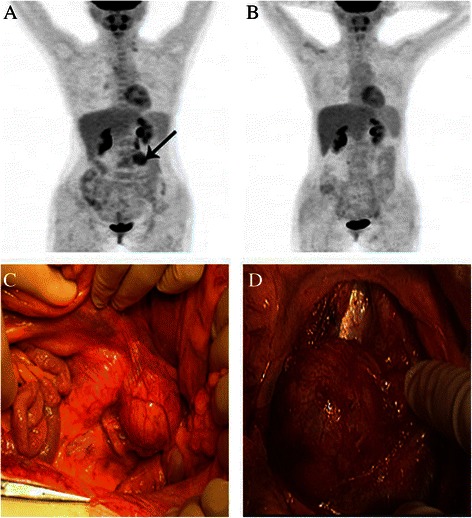
[[File:PET-CT scan.jpg|400px|thumb|none|PET-CT scan and macroscopic findings in a recurrent patient (case 11). (A) PET-CT scan before treatment; a black arrow points to the metastatic tumor in the left portion of the fourth lumbar vertebra. (B) PET-CT scan after treatment. (C) and (D) show the retroperitoneal tumor fused by several para-aortic lymph node,Qian Q, You Y, Yang J, et al. Management and prognosis of patients with ovarian sex cord tumor with annular tubules: a retrospective study. BMC Cancer. 2015;15:270. Published 2015 Apr 12. doi:10.1186/s12885-015-1277-y,https://www.ncbi.nlm.nih.gov/pmc/articles/PMC4408581/]] | [[File:PET-CT scan.jpg|400px|thumb|none|PET-CT scan and macroscopic findings in a recurrent patient (case 11). (A) PET-CT scan before treatment; a black arrow points to the metastatic tumor in the left portion of the fourth lumbar vertebra. (B) PET-CT scan after treatment. (C) and (D) show the retroperitoneal tumor fused by several para-aortic lymph node,Qian Q, You Y, Yang J, et al. Management and prognosis of patients with ovarian sex cord tumor with annular tubules: a retrospective study. BMC Cancer. 2015;15:270. Published 2015 Apr 12. doi:10.1186/s12885-015-1277-y,https://www.ncbi.nlm.nih.gov/pmc/articles/PMC4408581/]] | ||
| Line 16: | Line 17: | ||
{{WH}} | {{WH}} | ||
{{WS}} | {{WS}} | ||
[[Category: | [[Category:Disease]] | ||
[[Category:Types of cancer]] | |||
[[Category:Gynecology]] | |||
[[Category:Up-To-Date]] | |||
[[Category:Oncology]] | |||
[[Category:Medicine]] | |||
[[Category:Gynecology]] | |||
[[Category:Surgery]] | |||
[[Category:Radiology]] | |||
Latest revision as of 02:12, 6 May 2019
|
Sexcord/ stromal ovarian tumors Microchapters |
|
Differentiating Sexcord/ Stromal Ovarian Tumors from other Diseases |
|---|
|
Diagnosis |
|
Treatment |
|
Case Studies |
|
Sexcord/ stromal ovarian tumors other imaging findings On the Web |
|
American Roentgen Ray Society Images of Sexcord/ stromal ovarian tumors other imaging findings |
|
FDA on Sexcord/ stromal ovarian tumors other imaging findings |
|
CDC on Sexcord/ stromal ovarian tumors other imaging findings |
|
Sexcord/ stromal ovarian tumors other imaging findings in the news |
|
Blogs on Sexcord/ stromal ovarian tumors other imaging findings |
|
Risk calculators and risk factors for Sexcord/ stromal ovarian tumors other imaging findings |
Editor-In-Chief: C. Michael Gibson, M.S., M.D. [1]; Associate Editor(s)-in-Chief: ; Maneesha Nandimandalam, M.B.B.S.[2]
Overview
PET-CT may be helpful in the diagnosis of sexcord/ stromal ovarian tumors. On fluorodeoxyglucose-positron emission tomography/computed tomography malignant ovarian tumors generally have intense uptake, whereas tumors with a small solid content show decreased uptake.
Other Imaging Findings
- PET-CT may be helpful in the diagnosis sexcord/ stromal ovarian tumors
- On fluorodeoxyglucose-positron emission tomography/computed tomography malignant ovarian tumors generally have intense uptake, whereas tumors with a small solid content show decreased uptake. [1][2][3][4][5]

References
- ↑ Qian Q, You Y, Yang J, Cao D, Zhu Z, Wu M, Chen J, Lang J, Shen K (April 2015). "Management and prognosis of patients with ovarian sex cord tumor with annular tubules: a retrospective study". BMC Cancer. 15: 270. doi:10.1186/s12885-015-1277-y. PMID 25886261.
- ↑ Bristow RE, Simpkins F, Pannu HK, Fishman EK, Montz FJ (April 2002). "Positron emission tomography for detecting clinically occult surgically resectable metastatic ovarian cancer". Gynecol. Oncol. 85 (1): 196–200. doi:10.1006/gyno.2002.6611. PMID 11925145.
- ↑ Tomimatsu T, Fukuda Y, Mimura K, Yoshino K, Kato H, Tsuboyama T, Hori Y, Kimura T (December 2016). "Intense fluorodeoxyglucose uptake by a benign sclerosing stromal tumor of the ovary". Taiwan J Obstet Gynecol. 55 (6): 893–894. doi:10.1016/j.tjog.2016.03.005. PMID 28040143.
- ↑ Matsutani H, Nakai G, Yamada T, Yamamoto K, Ohmichi M, Narumi Y (December 2018). "Diversity of imaging features of ovarian sclerosing stromal tumors on MRI and PET-CT: a case report and literature review". J Ovarian Res. 11 (1): 101. doi:10.1186/s13048-018-0473-1. PMC 6302382. PMID 30572921.
- ↑ Kitajima K, Ueno Y, Maeda T, Murakami K, Kaji Y, Kita M, Suzuki K, Sugimura K (November 2011). "Spectrum of fluorodeoxyglucose-positron emission tomography/computed tomography and magnetic resonance imaging findings of ovarian tumors". Jpn J Radiol. 29 (9): 605–8. doi:10.1007/s11604-011-0610-x. PMID 21956364.