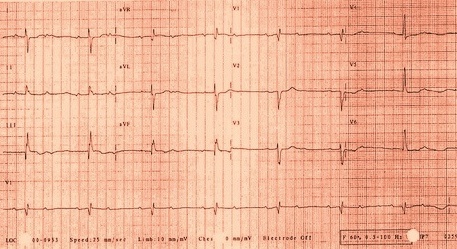
Second degree AV block electrocardiogram
|
Second degree AV block Microchapters |
|
Diagnosis |
|---|
|
Treatment |
|
Case Studies |
|
Second degree AV block electrocardiogram On the Web |
|
American Roentgen Ray Society Images of Second degree AV block electrocardiogram |
|
Risk calculators and risk factors for Second degree AV block electrocardiogram |
Editor-In-Chief: C. Michael Gibson, M.S., M.D. [1]; Associate Editor(s)-in-Chief: Cafer Zorkun, M.D., Ph.D. [2] Syed Musadiq Ali M.B.B.S.[3]
Electrocardiogram
Electrocardiographic Findings
Type I Second Degree AV Block
- Also called the Wenckebach phenomenon or Mobitz type I block[1][2]
- Intermittent failure of the supraventricular impulse to be conducted to the ventricles, not every P wave is followed by a QRS
- There is progressive prolongation of the PR interval until a P wave is blocked
- Progressive shortening of the RR interval until a P wave is blocked
- The RR interval containing the blocked P wave is shorter than the sum of 2 PP intervals[3]
- The increase in the PR interval is longest in the second conducted beat after the pause
- These rules may not be followed because of fluctuation in vagal tone and secondary to sinus arrhythmia.[4]
- In patients with normal QRS width, the block is usually located in the AV node
- There is progressive prolongation of the AH interval until the blocked P wave occurs
- When it is associated with bundle branch block, the block may occur in the AV node, His bundle or the contralateral bundle branch
- In 75% the block is in the AV node
- In 25% it is infranodal
Shown below is a two lead rhythm strip from a patient in the emergency room. The rhythm is sinus with second degree A/V block. Note the progressive lengthening of the PR interval and that the interval that brackets the blocked P wave is less than twice that of the RR interval. This recording suggests a Mobitz I A/V block, but some care has to be taken as the QRS that ends the pause in at least one case looks like a nodal escape beat.

Copyleft image obtained courtesy of ECGpedia, http://en.ecgpedia.org/wiki/Main_Page
Shown below is an image of an electrocardiogram showing type I second degree AV block (Wenckebach).

Copyleft image obtained courtesy of ECGpedia,http://en.ecgpedia.org/wiki/Main_Page
Type II Second-Degree AV Block (Mobitz Type II Block)
- There are intermittent blocked P waves
- In the conducted beats, the PR intervals remain constant
- The PR is fairly constant except that slight shortening may occur in the first beat after the blocked cycle. This is the result of improved conduction following the block
- Most patients with type II second-degree AV block have associated bundle branch block.
- In these instances the block is usually located distal to the His bundle, in approximately 27 to 35% of patients however, the lesion is located in the His bundle itself, and a narrow complex may be inscribed.
- 2:1 AV Block:
- Impossible to determine whether the second-degree AV block is type I or type II.
- A long rhythm strip is helpful to document any change in the behavior of the conduction ratio
- When the atrial rate is increased by exercise or by atropine, the AV block in type I tends to decrease and that in type II tends to increase
Shown below is an electrocardiogram of a 12 lead EKG with a 2:1 AV block.

Copyleft image obtained courtesy of ECGpedia, http://en.ecgpedia.org/wiki/Main_Page
Shown below is an electrocardiogram of a type II second degree AV block (Mobitz type II).

Copyleft image obtained courtesy of ECGpedia, http://en.ecgpedia.org/wiki/Main_Page
For more EKG examples of Second Degree AV Block click here
References
- ↑ Kerola T, Eranti A, Aro AL, Haukilahti MA, Holkeri A, Junttila MJ, Kenttä TV, Rissanen H, Vittinghoff E, Knekt P, Heliövaara M, Huikuri HV, Marcus GM (May 2019). "Risk Factors Associated With Atrioventricular Block". JAMA Netw Open. 2 (5): e194176. doi:10.1001/jamanetworkopen.2019.4176. PMC 6632153 Check
|pmc=value (help). PMID 31125096. - ↑ Trappe HJ (September 2016). "[Consciousness disorders from cardiological view]". Dtsch. Med. Wochenschr. (in German). 141 (19): 1361–9. doi:10.1055/s-0042-103177. PMID 27642736.
- ↑ Nelson WP (March 2016). "Diagnostic and Prognostic Implications of Surface Recordings from Patients with Atrioventricular Block". Card Electrophysiol Clin. 8 (1): 25–35. doi:10.1016/j.ccep.2015.10.031. PMID 26920166.
- ↑ Barold SS (March 2018). "Type I Wenckebach second-degree AV block: A matter of definition". Clin Cardiol. 41 (3): 282–284. doi:10.1002/clc.22874. PMC 6489887. PMID 29460961.