Retinoblastoma differential diagnosis

Jump to navigation Jump to search

Editor-In-Chief: C. Michael Gibson, M.S., M.D. [1]; Associate Editor(s)-in-Chief: Simrat Sarai, M.D. [2] Sahar Memar Montazerin, M.D.[3]

Overview

Differential diagnosis

Retinoblastoma must be differentiated from other diseases that cause leukocoria. Differential diagnosis of leukocoria in children include:


Leukocoria


Tumors


Congenital malformations


Vascular diseases


Inflammatory diseases


Trauma


Retinoblastoma
Medulloepithelioma
Leukemia
Combined retinal hamartoma
Astrocytic hamartoma (Bourneville’s tuberous sclerosis)


Persistent fetal vasculature (PFV)
Posterior coloboma
Retinal fold
Myelinated nerve fibers
Morning glory syndrome
Retinal dysplasia
Norrie’s disease
Incontinentia pigmenti
Cataract


Retinopathy of prematurity (ROP)
Coats’ disease
Familial exudative vitreoretinopathy (FEVR)


Ocular toxocariasis
Congenital toxoplasmosis
Congenital cytomegalovirus retinitis
Herpes simplex retinitis
Other types of fetal iridochoroiditis
Endophthalmitis


Intraocular foreign body
Vitreous hemorrhage
Retinal detachment
 
The above table adopted from Clinical Ophthalmic Oncology book [1]

Differential diagnosis of leukocoria

Disease/Condition Clinical presentation Demographics/History Diagnosis Other notes
Retinoblastoma[2][3]
  • Sporadic in 90% of the cases
  • The median age of diagnosis is 18 months
  • Bilateral in 70% of the cases
Coats'disease[4][5]
  • Yellowish appearance of leukocoria
  • P/E:exudative retinal detachment with vascular tortuosity and telangiectasia
  • +/- neurovascular glaucoma
  • Absence of calcification
  • Sporadic in 100% of the cases
  • Almost always unilateral
  • More common among boys
  • The median age of diagnosis 5 to 9 years
  • P/E is diagnostic in most of the cases
  • Ultrasound examination:
    • Complete retinal detachment
    • Absence of calcification
    • Exudative, mobile lipid material under retina
  • Fluorescein angiographyr reveals characteristic telangiectasias of small to medium-sized retinal vessels
Persistent fetal vasculature (formerly known as persistent hyperplastic primary vitreous)[5]
  • Presence of leukocoria in infancy which commonly accompanied with microphthalmia
  • presence of retrolental fibrovascular
  • +/- secondary cataract
  • Sporadic in the majority of cases
  • Always congenital (present at birth)
  • Rarely bilateral
  • P/E:microphthalmia and intraocular pressure
  • presence of elongated ciliary processes contracting into the retrolental mass
  • Ultrasound imaging:
    • Vitreous band from lens to optic nerve
    • Short axial length of eyes
  • Bilateral cases has been accompanied with protein C deficiency
Astrocytic hamartoma[1]
  • Presence of gray-yellow or translucent tumors involving the posterior pole near optic nerve
  • Presents at any age
  • Some has been associated with neurofibromatosis type 1/tuberous sclerosis
  • P/E:a sessile shape tumor and arising from the inner aspect of the sensory retina
  • Presence of small areas of calcification/complete calcification in older patients
  • Reticular pattern of fine blood vessels in fluorescein angiography
Retinopathy of prematurity (ROP)[1]
  • Absence of calcification
  • Presence of retinal contraction in one or both eyes
  • History of:
    • Prematurity (<32 weeks gestation)
    • Low birth weight (<1.5 kg)
    • Oxygen supplementation
  • Leukocoria is the late presentation of the disease
  • Always bilateral
  • P/E:Bilateral retinal avascularity and nonperfusion in temporal peripheral retina with fibrovascular proliferation in advanced cases
  • Ultrasound imaging:
    • Retinal detachment with retinal bands
  • Short axial length of eyes
Ocular toxocariasis [1]
  • Presence of retinal and/or vitreous traction in approximately all the cases
  • Presents at any age
  • Mostly unilateral
  • Ingestion of larvae leads to the infection
  • P/E:presence of granuloma and retinal traction
  • Ultrasound imaging:
    • Peripheral mass
    • Vitreoretinal band
    • Traction retinal detachment
  • Presence of eosinophils in the anterior chamber tap
  • Classified into three subtypes:
    • Macular granuloma
    • Peripheral granuloma
    • Endophthalmitis
Hereditary retinal syndrome
  • Generally causes vitreoretinal traction and ocular inflammation not seen in retinoblastoma
  • Fundus examination reveals the characteristic signs


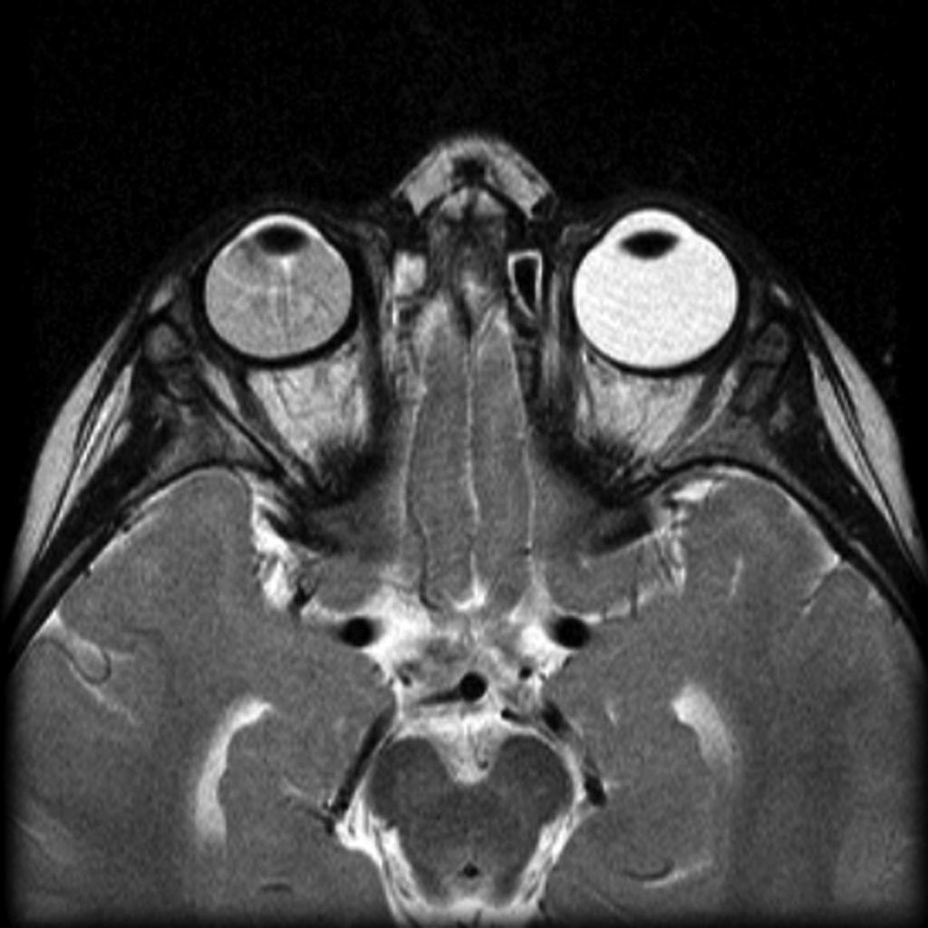
MRI of the orbit showing Coats disease - Case courtesy of Dr Michael Sargent, https://radiopaedia.org/. From the case https://radiopaedia.org/cases/6089
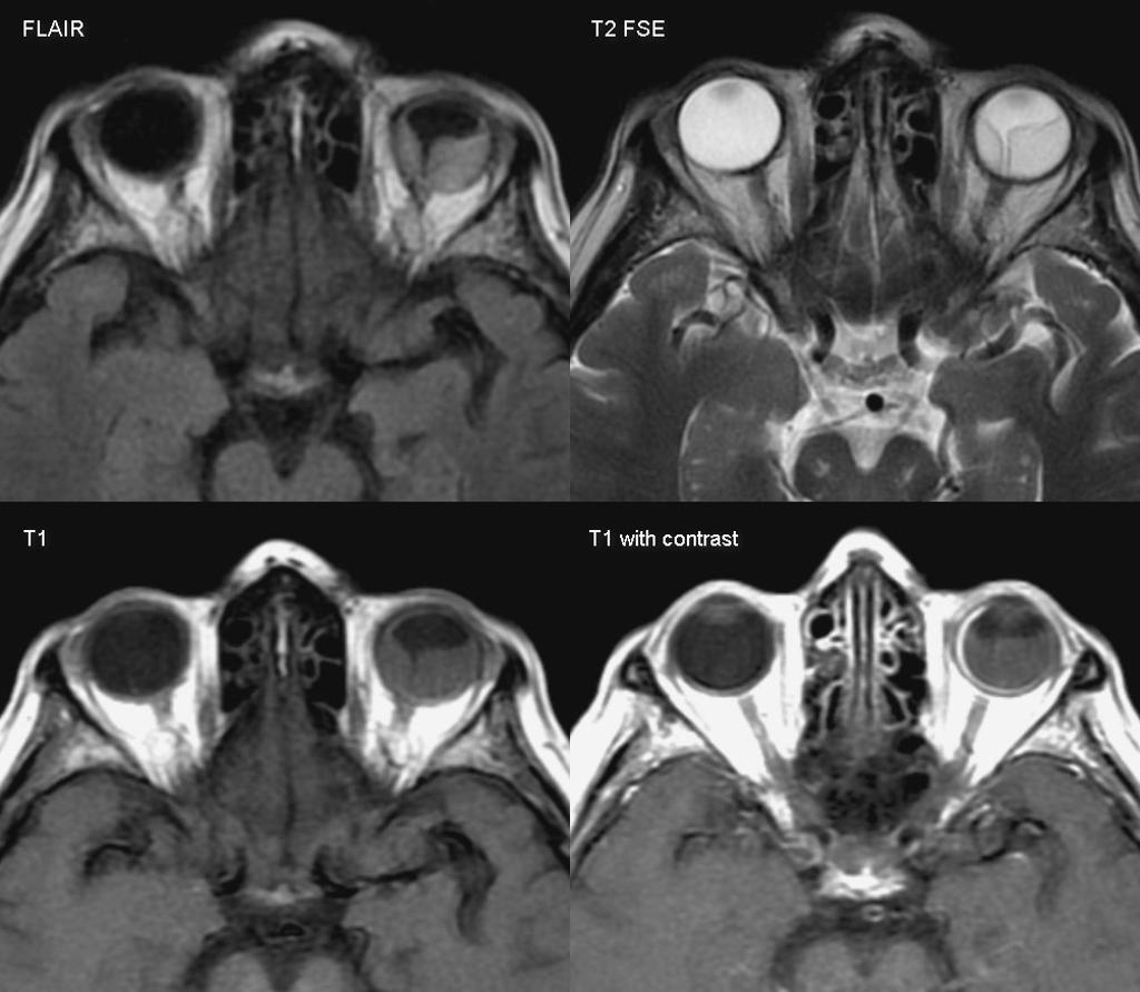
MRI of the orbit showing retinal detachment - Case courtesy of A.Prof Frank Gaillard, https://radiopaedia.org/. From the case https://radiopaedia.org/cases/3134
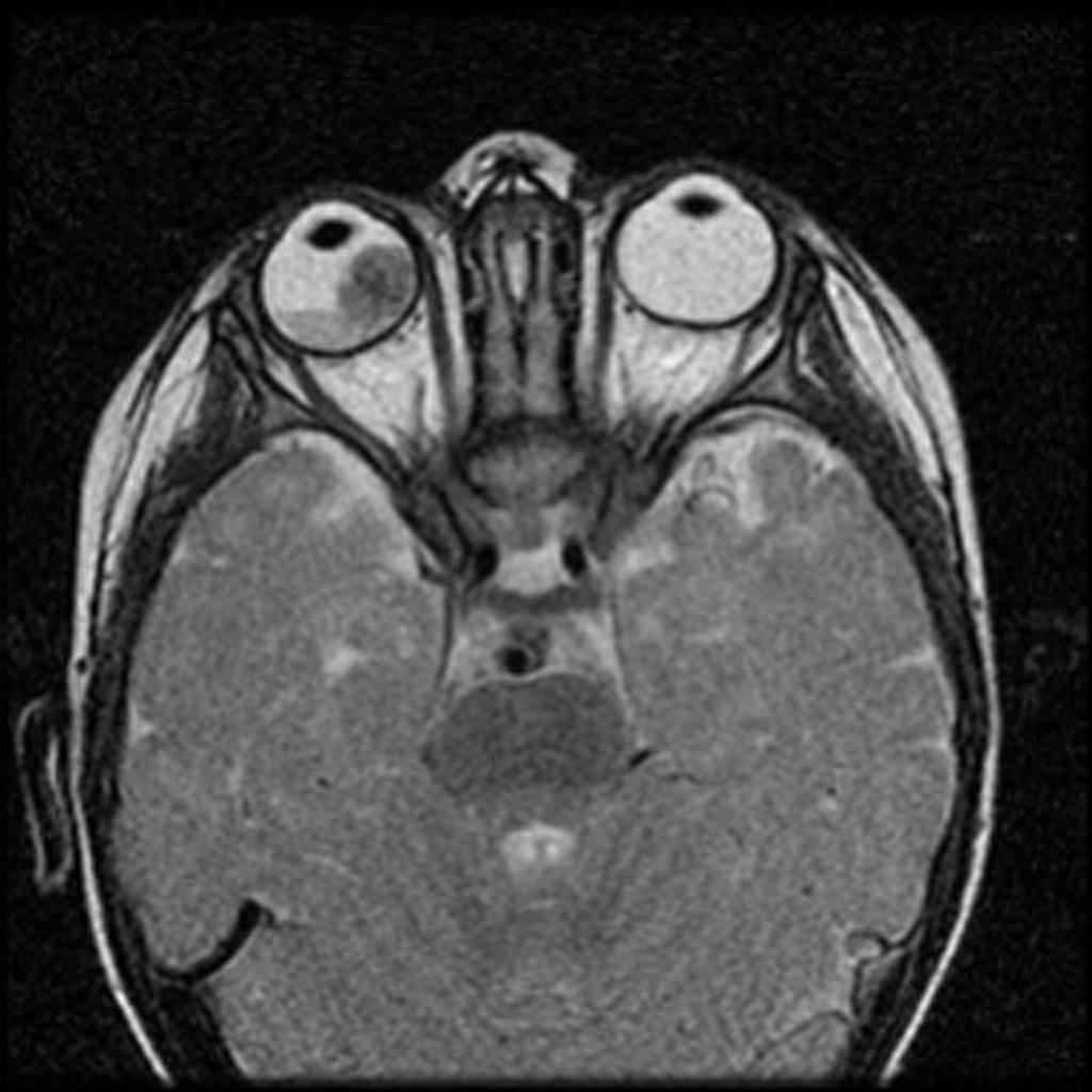
MRI of the orbit showing retinoblastoma - Case courtesy of https://radiopaedia.org/. From the case https://radiopaedia.org/cases/11877
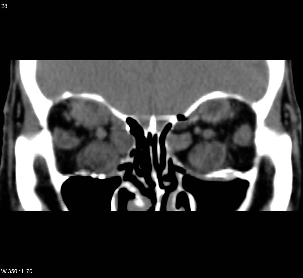
CT head showing hyperthyroid-induced orbitopathy - Case courtesy of A.Prof Frank Gaillard, https://radiopaedia.org/. From the case https://radiopaedia.org/cases/4854

References

  1. 1.0 1.1 1.2 1.3 Singh, Arun (2015). Clinical ophthalmic oncology : retinoblastoma. Heidelberg: Springer. ISBN 978-3-662-43451-2.
  2. Butros LJ, Abramson DH, Dunkel IJ (March 2002). "Delayed diagnosis of retinoblastoma: analysis of degree, cause, and potential consequences". Pediatrics. 109 (3): E45. PMID 11875173.
  3. Sachdeva R, Schoenfield L, Marcotty A, Singh AD (June 2011). "Retinoblastoma with autoinfarction presenting as orbital cellulitis". J AAPOS. 15 (3): 302–4. doi:10.1016/j.jaapos.2011.02.013. PMID 21680213.
  4. Silva RA, Dubovy SR, Fernandes CE, Hess DJ, Murray TG (December 2011). "Retinoblastoma with Coats' response". Ophthalmic Surg Lasers Imaging. 42 Online: e139–43. doi:10.3928/15428877-20111208-04. PMID 22165951.
  5. 5.0 5.1 Gupta N, Beri S, D'souza P (June 2009). "Cholesterolosis Bulbi of the Anterior Chamber in Coats Disease". J Pediatr Ophthalmol Strabismus. doi:10.3928/01913913-20090616-04. PMID 19645389.